- COVID-19 Tracker
- Biochemistry
- Anatomy & Physiology
- Microbiology
- Neuroscience
- Animal Kingdom
- NGSS High School
- Latest News
- Editors’ Picks
- Weekly Digest
- Quotes about Biology


Endosymbiotic Theory
Reviewed by: BD Editors
Endosymbiotic Theory Definition
Endosymbiotic theory is the unified and widely accepted theory of how organelles arose in organisms, differing prokaryotic organisms from eukaryotic organisms. In endosymbiotic theory, consistent with general evolutionary theory, all organisms arose from a single common ancestor. This ancestor probably resembled a bacteria, or prokaryote with a single strand of DNA surrounded by a plasma membrane. Throughout time, these bacteria diverged in form and function. Some bacteria acquired the ability to process energy from the environment in novel ways. Photosynthetic bacteria developed the pathways that enabled the production of sugar from sunlight. Other organisms developed novel ways to use this sugar is oxidative phosphorylation , which produced ATP from the breakdown of sugar with oxygen. ATP can then be used to supply energy to other reactions in the cell.
Both of these novel pathways led to organisms that could reproduce at a higher rate than standard bacteria. Other species, not being able to photosynthesis sugars or break them down through oxidative phosphorylation, decreased in abundance until they developed a novel adaptation of their own. The ability of endocytosis , or to capture other cells through the enfolding of the plasma membrane, is thought to have evolved around this time. These cells now had the ability to phagocytize , or eat, other cells. In some cells, the bacteria that were ingested were not eaten, but utilized. By providing the bacteria with the right conditions, the cells could benefit from their excessive production of sugar and ATP. One cell living inside of another is called endosymbiosis if both organisms benefit, hence the name of the theory. Endosymbiotic theory continues further, stating that genes can be transferred between the host and the symbiont throughout time.
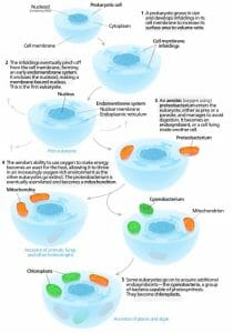
This gives rise to the final part of endosymbiotic theory, which explains the variable DNA and double membranes found in various organelles in eukaryotes. While the majority of cell products start in the nucleus, the mitochondria and chloroplast make many of their own genetic products. The nucleus, chloroplasts, and mitochondria of cells all contain DNA of different types and are also surrounded by double membranes, while other organelles are surrounded by only one membrane. Endosymbiotic theory postulates that these membranes are the residual membranes from the ancestral bacterial endosymbiont. If a bacteria was engulfed via endocytosis, it would be surrounded by two membranes. The theory states that these membranes survived evolutionary time because each organism retained the maintenance of its membrane, even while losing other genes entirely or transferring them to the nucleus. Endosymbiotic theory is supported by a large body of evidence. The general process can be seen in the following graphic.
Endosymbiotic Theory Evidence
The most convincing evidence supporting endosymbiotic theory has been obtained relatively recently, with the invention of DNA sequencing. DNA sequencing allows us to directly compare two molecules of DNA, and look at their exact sequences of amino acids. Logically, if two organism share a sequence of DNA exactly, it is more likely that the sequence was inherited through common descent than the sequence arose independently. If two unrelated organisms need to complete the same function, the enzyme they evolve does not have to look the same or be from the same DNA to fill the same role. Thus, it is much more likely that organisms who share sequences of DNA inherited them from an ancestor who found them useful.
This can be seen when analyzing the mitochondrial DNA (mtDNA) and chloroplast DNA of different organisms. When compared to known bacteria, the mtDNA from a wide variety of organisms contains a number of sequences also found in Rickettsiaceae bacteria. Fitting with endosymbiotic theory, these bacteria are obligate intracellular parasites. This means they must live within a vesicle of an organism that engulfs them through endocytosis. Like bacterial DNA, mtDNA and the DNA in chloroplasts is circular. Eukaryotic DNA is typically linear. The only genes missing from the mtDNA and those of the bacteria are for nucleotide, lipid, and amino acid biosynthesis. An endosymbiotic organism would lose these functions over time, because they are provided for by the host cell.
Further analysis of the proteins, RNA and DNA left in organelles reveals that some of it is too hydrophobic to cross the external membrane of the organelle, meaning the gene could never get transferred to the nucleus, as the cell would have no way of importing certain hydrophobic proteins into the organelle. In fact, chloroplasts and mitochondria have their own genetic code, and their own ribosomes to produce proteins. These proteins are not exported from the mitochondria or chloroplasts, but are needed for their functions. The ribosomes of mitochondria and chloroplasts also resemble the smaller ribosomes of bacteria, and not the large eukaryotic ribosomes. This is more evidence that the DNA originated inside of the organelles, and is separate completely from the eukaryotic DNA. This is consistent with endosymbiotic theory.
Lastly, the position and structure of these organelles lends to the endosymbiotic theory. The mitochondria, chloroplasts, and nuclei of cells are all surrounded in double membranes. All three contain their DNA in the center of the cytoplasm, much like bacterial cells. Although less evidence exists linking the nucleus to any kind of extant species, both chloroplasts and mitochondria greatly resemble several species of intracellular bacteria, existing in much the same manner. The nucleus is thought to have arisen through enfolding of the cell membrane, as seen in the graphic above. Throughout the world, there are various endosymbiont bacteria, all of which live inside other organisms. Bacteria exist almost everywhere, from the soil to inside our gut. Many have found unique niches within the cells of other organisms, and this is the basis of endosymbiotic theory.
Related Biology Terms
- Endosymbiont – An organism that lives with another organism, cause both organisms to receive benefits.
- Cyanobacteria – Still extant, cyanobacteria are photosynthetic bacteria whose ancestors probably became the chloroplasts of plant cells.
- Proteobacteria – The bacterial ancestor to the mitochondria organelle.
- Eukaryote – An organism with membrane bound organelles, thought to have evolved from endosymbiotic interactions.
Cite This Article
Subscribe to our newsletter, privacy policy, terms of service, scholarship, latest posts, white blood cell, t cell immunity, satellite cells, embryonic stem cells, popular topics, photosynthesis, horticulture, endocrine system, mitochondria, scientific method.

- Why Does Water Expand When It Freezes
- Gold Foil Experiment
- Faraday Cage
- Oil Drop Experiment
- Magnetic Monopole
- Why Do Fireflies Light Up
- Types of Blood Cells With Their Structure, and Functions
- The Main Parts of a Plant With Their Functions
- Parts of a Flower With Their Structure and Functions
- Parts of a Leaf With Their Structure and Functions
- Why Does Ice Float on Water
- Why Does Oil Float on Water
- How Do Clouds Form
- What Causes Lightning
- How are Diamonds Made
- Types of Meteorites
- Types of Volcanoes
- Types of Rocks
The Endosymbiotic Theory
The endosymbiotic theory is a scientific theory that proposes that some of the organelles in the eukaryotic cells, such as mitochondria and chloroplast , have originated from free-living prokaryotes ( bacteria and archaea ). Endosymbiosis is the relationship between two organisms when one lives within the other organism, eventually benefiting both partners.
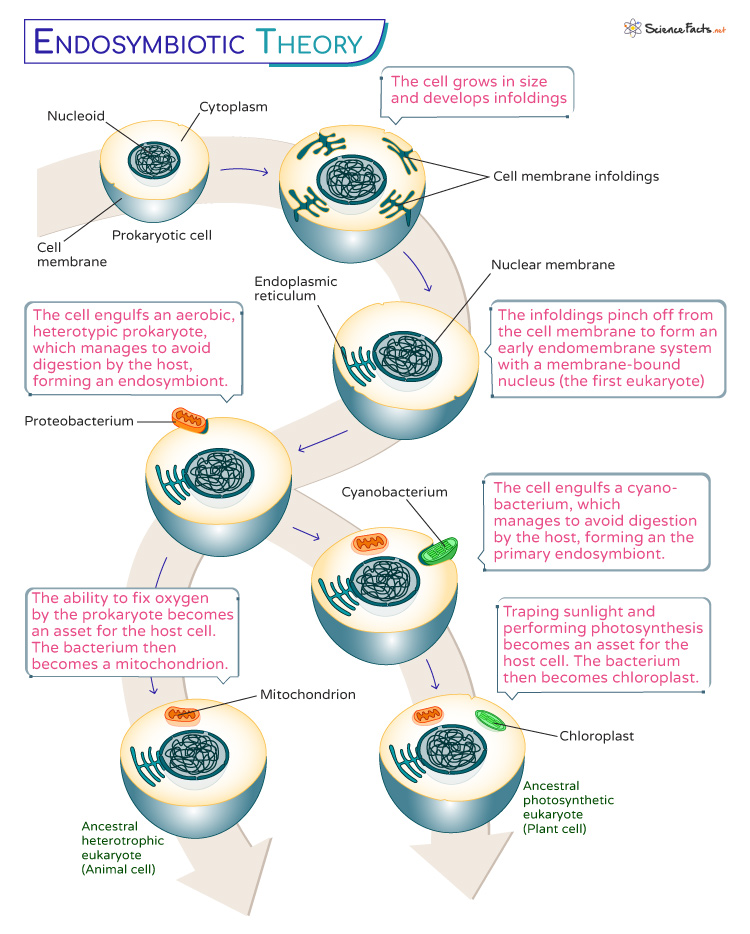
The endosymbiotic theory explains that when one organism, typically a microbe, takes up residence within the cell of another organism, over time, they form a close relationship that can be advantageous for both partners. The larger host cell provides a protected environment and essential nutrients. At the same time, the internalized microbe contributes its specialized functions, often becoming an organelle within the host cell.
The theory was conceptualized first by Konstantin Mereschkowsk in 1905 and then supported with evidence by Lynn Margulis in 1967.
The Theory of Endosymbiosis in Timeline
The German botanist Heinrich Anton de Bary coined the term ‘Symbiose’ to designate this coexistence. The concept of symbiosis , that two different organisms stably coexist and even give rise to a new type of organism, is attributed to Simon Schwendener.
19th Century
- In 1905, Russian biologist Konstantin Mereschkowski suggested that the origin of eukaryotic cells involved the engulfment of smaller prokaryotic cells.
- Around the same time, German botanist Andreas Schimper proposed that chloroplasts in plant cells might have originated from independent photosynthetic organisms.
1960s-1970s
- In 1967, Lynn Margulis proposed that eukaryotic cells, with their complex structures and organelles, resulted from symbiotic relationships between ancestral prokaryotic cells. He argued that mitochondria, the cellular powerhouses, were once free-living bacteria capable of aerobic respiration . Similarly, she suggested that chloroplasts originated from photosynthetic bacteria that became incorporated into host cells.
1980s-1990s
- Researchers discovered striking similarities between the DNA of organelles like mitochondria and chloroplasts and the DNA of modern-day bacteria.
- Protists like single-celled organisms are found to host certain bacteria fixing nitrogen (nitrogen-fixing bacteria).
2000s-Present
- In the 21st Century, endosymbiosis forms the basis of the origin of mitochondria, chloroplast, and other organelles. It also explains the development of complex multicellular organisms and the coevolution of symbiotic partners.
The Process of Endosymbiosis
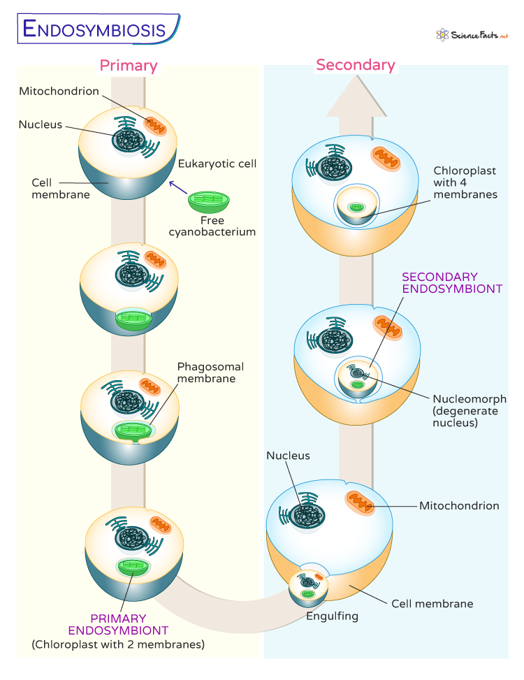
According to the endosymbiotic theory, the symbiotic origin of eukaryotic cells is a multi-event process.
1. Primary Endosymbiosis
It refers to the initial engulfment of a free-living bacterium by a host cell, creating a new organelle within the host cell. The most prominent examples of primary endosymbiosis are the origin of mitochondria and chloroplasts; both were once free-living cells. Here, two membranes surround the organelles; the inner is obtained from the bacterium, and the outer is derived from the host cell.
- Origin of Mitochondria : A eukaryotic cell engulfed a bacterium capable of aerobic respiration. This bacterium provides a valuable energy source through respiration. Over time, the host cell and the engulfed bacterium developed a mutually beneficial relationship. The bacterium became the mitochondrion, retaining its own DNA and membrane structure while working in tandem with the host cell.
- Origin of Chloroplasts : Here, an ancestral host cell captured a photosynthetic bacterium, which could convert sunlight into energy through photosynthesis . As with mitochondria, a symbiotic partnership formed, with the photosynthetic bacterium evolving into the chloroplast. It allowed the host cell to harness the power of photosynthesis for energy production.
2. Secondary Endosymbiosis
It involves a eukaryotic cell engulfing another eukaryotic cell already undergoing primary endosymbiosis. This secondary engulfment results in a more complex cellular arrangement, leading to the diversification of eukaryotic lineages and the emergence of new types of organelles.
- Formation of Plastids : These organelles involved in photosynthesis are found in various algae and plants. Different groups of algae have acquired plastids through secondary endosymbiosis, which consists of the engulfment of photosynthetic eukaryotic cells. In contrast to the two membranes of primary organelles, four membranes surround chloroplasts obtained by secondary endosymbiosis. In most cases, the nucleus of the engulfed cell disappears, with the remains of this nucleus still found lying between the two pairs of membranes. This structure is called a nucleomorph.
Thus, the endosymbiotic theory explains the presence of double-membraned organelles within protists.
Evidence that Supports the Endosymbiotic Theory
There are several proofs to support the Endosymbiotic Theory. However, the discovery of independent DNA (from the host) in mitochondria and chloroplasts supported the theory the most. The other evidences are as follows:
- Structural Similarities : Mitochondria and chloroplasts share structural characteristics with free-living bacteria, such as double membranes and DNA. Both the organelles are almost of the same size as the bacterial cell.
- Reproduction : Mitochondria and chloroplasts replicate within the cell independently, similar to how bacteria reproduce.
- Genetic Evidence : The DNA within mitochondria and chloroplasts is more similar to bacterial DNA than the host cell’s nucleus.
- Evolutionary Relationships : Analysis of genetic sequences shows that mitochondria and chloroplasts are more closely related to specific groups of bacteria than eukaryotic cells.
However, the statement that mitochondria and chloroplasts are much larger than prokaryotic cells does not support the endosymbiotic theory.
What is the Importance of the Endosymbiotic Theory
It is important because the theory explains the origin of the eukaryotic cells. It also describes how chloroplast and mitochondria might have originated from once free-living prokaryotes. This understanding has reshaped our perception of how fundamental cellular organelles came to be and how multicellular life forms arose.
The endosymbiotic theory also highlights how cooperation and symbiosis have played pivotal roles in shaping cellular evolution.
- Endosymbiosis – Ib.bioninja.com.au
- Endosymbiosis and the Evolution of Eukaryotes – Bio.libretexts.org
- Endosymbiotic Theories forEukaryote Origin – Royalsocietypublishing.org
- Endosymbiosis – Evolution.berkeley.edu
- Endosymbiosis: The Feeling is Not Mutual – Ncbi.nlm.nih.gov
- Endosymbiosis – Cell.com
Article was last reviewed on Tuesday, October 3, 2023
Related articles
Leave a Reply Cancel reply
Your email address will not be published. Required fields are marked *
Save my name, email, and website in this browser for the next time I comment.
Popular Articles
Join our Newsletter
Fill your E-mail Address
Related Worksheets
- Privacy Policy
© 2024 ( Science Facts ). All rights reserved. Reproduction in whole or in part without permission is prohibited.
An official website of the United States government
The .gov means it’s official. Federal government websites often end in .gov or .mil. Before sharing sensitive information, make sure you’re on a federal government site.
The site is secure. The https:// ensures that you are connecting to the official website and that any information you provide is encrypted and transmitted securely.
- Publications
- Account settings
Preview improvements coming to the PMC website in October 2024. Learn More or Try it out now .
- Advanced Search
- Journal List
- Philos Trans R Soc Lond B Biol Sci
- v.370(1678); 2015 Sep 26
Endosymbiotic theories for eukaryote origin
For over 100 years, endosymbiotic theories have figured in thoughts about the differences between prokaryotic and eukaryotic cells. More than 20 different versions of endosymbiotic theory have been presented in the literature to explain the origin of eukaryotes and their mitochondria. Very few of those models account for eukaryotic anaerobes. The role of energy and the energetic constraints that prokaryotic cell organization placed on evolutionary innovation in cell history has recently come to bear on endosymbiotic theory. Only cells that possessed mitochondria had the bioenergetic means to attain eukaryotic cell complexity, which is why there are no true intermediates in the prokaryote-to-eukaryote transition. Current versions of endosymbiotic theory have it that the host was an archaeon (an archaebacterium), not a eukaryote. Hence the evolutionary history and biology of archaea increasingly comes to bear on eukaryotic origins, more than ever before. Here, we have compiled a survey of endosymbiotic theories for the origin of eukaryotes and mitochondria, and for the origin of the eukaryotic nucleus, summarizing the essentials of each and contrasting some of their predictions to the observations. A new aspect of endosymbiosis in eukaryote evolution comes into focus from these considerations: the host for the origin of plastids was a facultative anaerobe.
1. Introduction
Early evolution is an important part of life's history, and the origin of eukaryotes is certainly one of early evolution's most important topics, as the collection of papers in this special issue attests. There are various perspectives from which eukaryote origins can be viewed, including palaeontological evidence [ 1 ], energetics [ 2 ], the origin of eukaryote-specific traits [ 3 , 4 ] or the relationships of the different eukaryotic groups to one another [ 5 ]. This paper will look at eukaryote origins from the standpoint of endosymbiotic theory, and how different versions of endosymbiotic theory tend to square off with the data that we have for eukaryotic anaerobes and with regard to data from gene phylogenies. Endosymbiotic theory has a long and eventful history, virtuously summarized in Archibald's book [ 6 ], and speaking of history, here is a good place to dispel a myth—about Altmann.
One can occasionally read (though we will politely provide no examples) that Altmann [ 7 ] is to be credited with the idea of symbiotic theory for the origin of mitochondria, but that is incorrect. Those of us who can read German and who have a copy of Altmann's 1890 book can attest: in the 1890 book, Altmann was not interested in mitochondria, and he did not propose their symbiotic origin. He mentioned neither mitochondria (nor their older name, chondriosomes) nor endosymbiosis in his book on ‘bioblasts’. To Altmann, everything in eukaryotic cells consisted of bioblasts, including the cytosol, the nucleus and the chromosomes. His bioblasts corresponded to a chemical organization state of matter that was larger than the molecule but smaller than the cell ‘the smallest morphological unit of organized material’ (‘ die kleinste morphologische Einheit der organisirten Materie ’) [ 8 , p. 258]. They would maybe correspond in size roughly to what we today call macromolecular complexes, which however cannot be seen in the light microscopes of Altmann's day. He also distinguished autoblasts, cytoblasts, karyoblasts and somatoblasts, which are mentioned far less often than bioblasts. A scholarly treatise of Altmann in the context of symbiotic theory, and why he cannot be credited with having suggested endosymbiotic theory, can be found in Höxtermann & Mollenhauer [ 8 ].
The concept of symbiosis (Latin, ‘living together’), that two different organisms can stably coexist and even give rise to a new type of organism, traces to Simon Schwendener [ 9 ], a Swiss botanist who discovered that lichens consist of a fungus and a photosynthesizer. The German botanist Heinrich Anton de Bary (1878) coined the term ‘ Symbiose ’ to designate this type of coexistence [ 10 ]. Schimper [ 11 ] is sometimes credited with the discovery of endosymbiotic theory, but his treatise of the topic is wholly contained in a footnote that translates to this: ‘If it can be conclusively confirmed that plastids do not arise de novo in egg cells, the relationship between plastids and the organisms within which they are contained would be somewhat reminiscent of a symbiosis. Green plants may in fact owe their origin to the unification of a colorless organism with one uniformly tinged with chlorophyll’ [ 11 , pp. 112–113]. That was all he wrote on the possibility of symbiotic plastid origin. The sentence immediately following that one in Schimper's famous footnote, however, is also significant, as we will see in a later passage about Portier and the symbiotic origin of mitochondria; it translates to this: ‘According to Reinke (Allg. Botanik, p. 62) the chlorophyll bodies [Chlorophyllkörner, another name for plastids in Schimper's day] might even have the ability to live independently; he observed this phenomenon, as communicated to me, and published with kind permission, in a rotting pumpkin, the chloroplastids of which, surrounded by Pleosporahyphae, continued to vegetate in dead cells and multiplied by division’ [ 11 , p. 113]. Clearly, Reinke was observing the proliferation of contaminating bacteria, not of free-living organelles.
Schimper [ 11 , 12 ] did, however, champion the case that plastids proliferate through division. That was important for the Russian biologist Constantin Mereschkowsky, who probably delivered the first thoroughly argued case that some cells arose through the intracellular union of two different kinds of cells (endosymbiosis), in his 1905 paper [ 13 ] that has been translated into English [ 14 ]. Mereschkowsky [ 13 ] said three things: (i) plastids are unquestionably reduced cyanobacteria that early in evolution entered into a symbiosis with a heterotrophic host, (ii) the host that acquired plastids was itself the product of an earlier symbiosis between a larger, heterotrophic, amoeboid host cell and a smaller ‘micrococcal’ endosymbiont that gave rise to the nucleus, and (iii) the autotrophy of plants is an inheritance, in toto , from cyanobacteria [ 13 ].
Mereschkowsky's scheme was more fully elaborated but basically unchanged in his 1910 series [ 15 ]: there were two kinds of fungi, those that evolved a nucleus without endosymbiosis and those that once possessed plastids but became secondarily non-photosynthetic, today we call them the oomycetes, and there is still no consensus on the issue of whether they ever had plastids or not. The branches in Mereschkowsky's tree occasionally unite via endosymbiosis to produce fundamentally and radically new kinds of organisms (plants, for example) [ 15 , 16 ]. A more modern version of symbiosis in cell evolution would have to include the symbiotic origin of mitochondria, archaea and the concept of secondary endosymbiosis. Endosymbiotic theories have it that cells unite, one inside the other, during evolution to give rise to novel lineages at the highest taxonomic levels, via combination. That is not the kind of evolution that Darwin had in mind; his view of evolution was one of gradualism.
Many biologists still have a problem with the notion of endosymbiosis and hence prefer to envisage the origin of eukaryotes as the product of gene duplication, point mutation and micromutational processes [ 17 ]. A 2007 paper by the late Christian de Duve [ 18 ] is now often taken as the flagpole for micromutational theories of eukaryote origin, but de Duve, like the late Lynn Margulis [ 19 ], always categorically rejected the evidence that mitochondria and hydrogenosomes—anaerobic forms of mitochondria [ 20 , 21 ]—share a common ancestor. No anaerobic form of mitochondria ever fits into classical endosymbiotic theory. This is because classical (Margulis's version of) endosymbiotic theory [ 19 ] was based on the premise that the benefit of the endosymbiotic origins of mitochondria was founded in oxygen utilization, while de Duve's versions went one step further and suggested that even the endosymbiotic origin of peroxisomes was founded in oxygen utilization [ 18 ]. Anaerobic mitochondria were never mentioned and hydrogenosomes, if they were mentioned, were explained away as not being mitochondria [ 18 , 19 ]. The overemphasis of oxygen in endosymbiotic theory and how the focus on oxygen led to much confusion concerning the phylogenetic distribution and evolutionary significance of anaerobic forms of mitochondria has been dealt with elsewhere [ 22 – 24 ].
There is one alternative to classical endosymbiotic theory that took anaerobic mitochondria and hydrogenosomes into account, the hydrogen hypothesis [ 25 ]; it predicted (i) all eukaryotes to possess mitochondria or to have secondarily lost them, (ii) that the host for mitochondrial origins was an archaeon, the eukaryotic state having arisen in the wake of mitochondrial origins, and (iii) that aerobic and anaerobic forms should interleave on the eukaryotic tree. Though radical at the time, prediction (i) was borne out [ 26 – 29 ], and so was prediction (ii) [ 30 – 32 ], as well as (iii) [ 21 , 33 ]. Furthermore, only recently, it has been recognized that the invention of eukaryotic specific traits required more metabolic energy per gene than prokaryotes have at their disposal, and that mitochondria afforded eukaryotic cells an orders of magnitude increase in the amount of energy per gene, which (finally) explains why the origin of eukaryotes corresponds to the origin of mitochondria [ 2 , 34 ]. But there is more to eukaryote origins than just three predictions and energy. There is the origin of the nucleus to deal with [ 35 ], and the role that gene phylogenies have come to play in the issues. In addition, there is the full suite of characters that distinguish eukaryotes from prokaryotes to consider (meiosis, mitosis, cell cycle, membrane traffic, endoplasmic reticulum (ER), Golgi, flagella and all the other eukaryote-specific attributes, including a full-blown cytoskeleton—not just a spattering of prokaryotic homologues for cytoskeletal proteins [ 31 ]), but here our focus is on endosymbiotic theories, not the autogenous origin of ancestrally shared eukaryotic characters, whose origins for energetic reasons come in the wake of mitochondrial origin [ 34 ].
2. Gene trees, not as simple as it sounds
To get a fuller picture of eukaryote origins, we have to incorporate lateral gene transfer (LGT) among prokaryotes, endosymbiosis and gene transfer from organelles to the nucleus into the picture. That is not as simple as it might seem, because it has become apparent that individual genes have individual and differing histories. Thus, in order to get the big picture, we would have to integrate all individual gene trees into one summary diagram in such a way as to take the evolutionary affinities of the plastid (a cyanobacterium), the mitochondrion (a proteobacterium) and the host (an archaeon) into account. Nobody has done that yet, although there are some attempts in that direction [ 36 ]. In 2015, our typical picture of eukaryotic origins entails either a phylogenetic tree based on one gene or, more commonly now, a concatenated analysis of a small sample of genes (say 30 or so from each genome), which generates a tree, the hope being that the tree so obtained will be representative for the genome as a whole and thus will have some predictive character for what we might observe in phylogenies beyond the 30 or so genes used to make the tree. The 30 or so genes commonly used for such concatenated phylogenies are mostly ribosomal proteins or other proteins involved in information processing, genes that Jim Lake called informational genes in 1998 [ 37 ].
But because of the role of endosymbiosis in eukaryote cell evolution, eukaryotes tend to have two evolutionarily distinct sets of ribosomes (archaeal ribosomes in the cytosol and bacterial ribosomes in the mitochondrion), or sometimes three (an additional bacterial set in the plastid [ 38 ]) and in rare cases four sets of active ribosomes (yet one more set in algae that possess nucleomorphs) [ 39 ]. The ‘core set of genes’ approach, in all of its manifestations so far, only queried cytosolic ribosomes for eukaryotes, and thus only looked at the archaeal component of eukaryotic cell history. Some of us have been worried that by looking only at genes that reflect the archaeal component of eukaryotic cells we might be missing a lot, because it was apparent early on that many genes in eukaryotes do not stem from archaea, but from bacteria instead and, most reasonably under endosymbiotic theory, from organelles [ 40 , 41 ].
An early study looking at the phylogeny of the core gene set, which largely but not entirely corresponds to the ribosomal protein superoperon of prokaryotes, came to the conclusion that the information contained within the alignment is problematic because of the low amount of sequence conservation involved across many of the sites [ 42 ]. Concerns were also voiced that the 30 genes of the set, if analysed individually, might not have the same history and that concatenation might thus be a problem [ 43 ], but that did not stop bioinformaticians [ 44 ] from rediscovering the same set of 30 or so genes and making a tree that looked remarkably similar to the rRNA tree in most salient aspects, in particular as regards the position of the eukaryotes. By that time it was reasonably well-known that the genes of archaeal origin in eukaryotes are not representative of the genomes as a whole; they constitute a minority of the genome and are vastly outnumbered by genes of bacterial origin [ 45 ]. Despite that, the attention in the issue of eukaryote origins has, with few exceptions [ 46 – 48 ], remained focused on the archaeal component, and it will probably stay that way until improved methods to summarize the information contained in thousands of trees come to the fore.
Always critical of the branches in trees that phylogenetic methods produce [ 49 ], Embley and colleagues looked at the conserved core set with more discerning phylogenetic methods [ 30 , 50 , 51 ] and found that the archaeal component of eukaryotes branches within the archaea. These new trees tend to group the eukaryotes with the crenarchaeotes, specifically with the TACK superphylum of archaea [ 31 ], while at the same time tending to locate the root of the archaea among the euryarchaeotes, sometimes among the methanogens [ 52 ].
Now is a good time to have a look at endosymbiotic theories and related ideas for the origin of eukaryotes, their nucleus and their mitochondria. In doing so, we pick up on our own earlier reviews of the topic [ 22 , 53 ], the figures of which have become popular [ 31 ]. In the next section, we summarize what various models say, starting with models for the origin of the nucleus, and then move on to models for the origins of chloroplasts and mitochondria.
3. The nucleus
The nucleus is a defining feature of eukaryotes [ 54 ]. Theories for the evolution of the nucleus are usually based (i) on invaginations of the plasma membrane in a prokaryote or (ii) on endosymbiosis of an archaeon in a eubacterial host or (iii) on an autogenous origin of a new membrane system including the nuclear envelope in a host of archaeal origin after acquisition of mitochondria. The endosymbiotic theory for the origin of the nucleus started with Mereschkowsky [ 13 ]. He postulated that the nucleus evolved from a prokaryote (mycoplasma), which was engulfed by an amoeboid cell homologous to the eukaryotic cytosol ( figure 1 a ; [ 15 ]).

Models describing the origin of the nucleus in eukaryotes. ( a – o ) Schematic of various models accounting for the origin of the nucleus. Archaeal cells/membranes are represented with red, while blue indicates eubacterial cells/membranes. Black membranes are used when the phylogenetic identity of the cell is not clear or not specified. See also [ 22 , 53 ].
Cavalier-Smith argued that nuclear and ER membranes originated through invaginations of the plasma membrane of a prokaryotic cell ( figure 1 b ; [ 55 – 58 ]). He suggested that the prokaryote initially lost its cell wall and thereby gained the ability to phagocytose food particles. Ribosomes, primarily attached to the plasma membrane, became internalized, but still attached to the membrane, resulting first in the rough ER and out of it the nuclear envelope. Gould & Dring [ 59 ] presented a different model in 1979 where they described that endospore formation of Gram-positive bacteria resulted in the origin of the nucleus. The protoplast of a single cell divides during endospore formation in such a manner that the cell engulfs a portion of its own cytoplasm, which than becomes surrounded by a double membrane resulting in the cell's nucleus ( figure 1 c ; [ 59 ]). In the 1990s, several models for the origin of the nucleus via endosymbiosis (sometimes called endokaryotic theories) were published, but only few refer to Mereschkowsky's original suggestion. They have in common that they envisage a eubacterial host that engulfed an archaebacterial endosymbiont that underwent a transformation into the nucleus ( figure 1 d ; [ 60 , 61 ]). Fuerst & Webb [ 62 ] observed that the DNA in the freshwater budding eubacterium Gemmata obscuriglobus (a member of the Planctomyces-Pirella group) appears to be surrounded by a folded membrane, the organization of which was thought to resemble the nucleus ( figure 1 e ; [ 62 ]). Later papers were less cautious and called this structure a nucleus outright [ 63 ]; subsequent work on Gemmata showed that the inner membrane is simply an invagination of the plasma membrane [ 64 ], as had been previously pointed out [ 53 ]. Searcy & Hixon [ 65 ] interpreted thermophilic acidophilic sulfur-metabolizing archaebacteria lacking a rigid cell wall but having a well-developed cytoskeleton as a primary stage for the evolution of eukaryotic cells ( figure 1 f ; [ 65 ]).
Lake & Rivera [ 66 ] suggested an endosymbiosis in which a bacterium engulfed an archaeon (crenarchaeon) for the origin of eukaryotes ( figure 1 g ). A vesicular model for the origin of the nucleus in a cell that had a mitochondrial endosymbiont was proposed ( figure 1 h ; [ 40 ]). It posits a role for gene transfer and the origin of bacterial lipids in the origin of the eukaryotic endomembrane system, and in a subsequent formulation [ 35 ] it posits a causal relationship between the origin of spliceosomes and the origin of nucleus–cytosol compartmentation (this aspect is discussed in more detail in a later section). Moreira & López-García [ 67 , 68 ] modified the endokaryotic model, invoking the principle of anaerobic syntrophy (H 2 -dependence) for the origin of the nucleus. They postulated a fusion of plasma membranes in an agglomeration of δ-proteobacteria entrapping a methanogenic archaebacterium, which evolved to the nucleus ( figure 1 i ; [ 67 , 68 ]). The kind of fusion of plasma membranes among free-living cells that Moreira & Lopez-Garcia [ 67 , 68 ] envisage has not been observed for bacteria, but it is known to occur among archaea [ 69 ]. Lynn Margulis presented another symbiogenic theory for the origin of the nucleus. She suggested a symbiosis between a spirochaete and an archaebacterium without a cell wall (most likely Thermoplasma -like in her view), leading to both the eukaryotic flagellum and the nucleus ( figure 1 j ; [ 19 , 70 ]). A viral origin for the nucleus involving poxviruses was suggested in 2001 by Bell in the context of syntrophic consortia involving methanogens ( figure 1 k ; [ 71 ]). Horiike postulated a model in which the nucleus emerged from an archaeal endosymbiont ( Pyrococcus -like), which was engulfed by a γ -proteobacterium ( figure 1 l ; [ 72 ]). An origin of eukaryotes (hence implicitly or explicitly their nucleus) prior to prokaryotes has also been repeatedly suggested (figure l m ; [ 73 – 75 ]). Penny argues that prokaryotes, which he and Forterre [ 73 ] sometimes call ‘akaryotes’ [ 75 ], arose from this eukaryote ancestor via Forterre's thermoreduction hypothesis—a transition to the prokaryotic state from a eukaryotic ancestor in response to higher temperatures.
More recently, the community of scientists interested in cytoskeletal evolution have—in unaltered form—rekindled Cavalier-Smith's hypothesis of an autogenous (non-symbiotic) origin of a phagocytosing amitochondriate eukaryote (an archezoon) via point mutational changes leading to a host that does not need a mitochondrion at all to enjoy its phagocytotic lifestyle, but acquires one nonetheless ( figure 1 n ; [ 76 ]).
Forterre [ 77 ] departed from thermoreduction and introduced a new variant of the endokaryotic hypothesis, one that got planctomycetes (a member of the PVC group: Planctomycetes, Verrucomicrobia, Chlamydiae) involved in eukaryote origin as the bacterial host for the engulfment of a thaumarchaeon as the nucleus, followed by invasions of retroviruses and nucleo-cytoplasmic large DNA viruses (NCLDV). In this theory, the PTV (for PVC–thaumarchaeon–virus) fusion hypothesis, the PVC bacterium provides universal components of eukaryotic membranes required also for the formation of the nucleus and the thaumarchaeon provides informational and operational proteins and precursors of the modern eukaryotic cytoskeleton and vesicle trafficking system ( figure 1 o ; [ 77 ]).
A problem with all models that envisage a role for planctomycetes in eukaryote origin is that there is no molecular phylogenetic evidence that would link any lineage of planctomycetes with eukaryotes [ 78 ]. The problems with theories that derive the nucleus from an endosymbiont are numerous and have been listed in detail elsewhere [ 40 ]; in essence, they fail to explain why the nuclear compartment is so fundamentally different from any free-living cell from the standpoints of (i) biosynthetic or ATP-generating physiology (altogether lacking in the nuclear compartment), (ii) membrane topology (no free-living cell is bounded similarly), (iii) permeability (no prokaryotic cytosol is contiguous with the environment via pores), and (iv) division (dissolution of a superficial homologue to the plasma membrane once per cell division in eukaryotes with open mitosis). Endosymbiotic theories for plastid and mitochondrial origin do not have those problems. A problem with the thermoreduction hypothesis is that it does not address the issue of where eukaryotes come from in the first place, it just takes their origin as a given. The recognition that the common ancestor of eukaryotes possessed a mitochondrion [ 30 , 32 , 79 ] is a severe problem for thermoreduction hypotheses, because the eukaryote has to first give rise to a prokaryote (the mitochondrial ancestor) that is required for its own origin, a sequence of events that, at face value, requires time to run backwards. Thermoreduction hypotheses are generally silent regarding the origin of mitochondria. Very few models for the origin of the nucleus, possibly only one, derive the nucleus in an archaeal host that possessed a mitochondrion. That model posits the nuclear membrane to arise from vesicles of membranes consisting of bacterial lipids [ 40 ] and invokes the need to separate splicing from translation as the selective pressure that led to the fixation of the compartmentation into nucleoplasm and cytoplasm [ 35 ].
The recent focus both on the evolution of cytoskeletal components [ 76 ] and on an autogenous (non-symbiotic) origin of a phagocytosing amitochondriate eukaryote point to a problem that should be mentioned. That theory, once called the archezoa hypothesis [ 55 , 56 ], now sometimes called the phagocytosing archaeon theory [ 31 ], envisages that point gradual changes lead to a prokaryotic host that can perform fully fledged eukaryotic phagocytosis (a quite complex process). These theories have it that phagocytosis is the key character that enabled the endosymbiotic origin of mitochondria. A problem common to those theories is that the phagocytotic, primitively amitochondriate eukaryote does not need a mitochondrion at all, and if there were some construable selective advantage then eukaryotes should have arisen from prokaryotes in multiple lineages independently. That has always been one of the weakest aspects of autogenous theories, in addition to the bioenergetic aspects [ 34 ].
4. The origin of mitochondria (and chloroplasts)
Endosymbiotic theory for the origin of chloroplasts and mitochondria started again with Mereschkowsky [ 13 ] and his idea about a symbiosis between ‘chromatophores’ (plastids) and a heterotrophic amoeboid cell. He contradicted the orthodox view that chromatophores are autogenous organs of the plant cells; he saw them as symbionts, extrinsic bodies or organisms, which entered into the host's plasma establishing a symbiotic relationship. The host for the origin of plastids itself originated, in his view, from an earlier symbiosis between a heterotrophic, amoeboid cell and a ‘micrococcal’ endosymbiont that gave rise to the nucleus ( figure 2 a ; [ 13 ]). Comparison of physiological and anatomic attributes of plastids and cyanobacteria known at that time led him to the certain conclusion that the endosymbionts were ‘cyanophyceae’ (cyanobacteria) that entered into symbioses with amoeboid or flagellated cells on several independent occasions, leading to a plant kingdom having several independent origins. That is, he viewed the different coloured plastids of algae (red, green, brown, golden) as inheritances from different endosymbionts, each having those different pigmentations. Although he was wrong on that specific interpretation—today there is broad agreement that the plastids of all plants and algae have a single origin [ 80 – 82 ]—he was right with the endosymbiotic, cyanobacterial origin of plastids.

Models describing the origin of mitochondria and/or chloroplasts in eukaryotes. ( a – q ) Schematic of various models accounting for the origin of mitochondria and/or chloroplasts. Archaeal cells/membranes are represented with red, while blue indicates eubacterial cells/membranes. Black membranes are used when the identity of the cell is not clear and green is used for cyanobacterial derived cells/membranes. See also [ 22 ].
Mereschkowsky failed, however, to recognize the endosymbiotic origin of mitochondria, although the physiological properties of cells that he explained with the endosymbiotic origin of the nucleus are, from today's perspective, properties of mitochondria [ 15 ]. As very readably explained by Archibald [ 6 ], Portier developed (in French) the idea that there was a close relationship between bacteria and mitochondria and that mitochondria were involved in numerous processes in the cell. But like Schimper in his footnote regarding plastids, which we translated above, Portier proposed that mitochondria could be cultured outside their host cells, and this precipitated unforgiving criticism from his contemporaries [ 6 ]. Clearly, both Reinke (as cited in Schimper's footnote that we translated above) and Portier were observing the proliferation of contaminating bacteria, not of free-living organelles. Wallin [ 83 ] developed the endosymbiotic theory further for mitochondria, in English. He recognized that these organelles are descendants of endosymbiotic bacteria, but it remained very unclear what his idea about the host was ( figure 2 b ; [ 83 ]). Like Portier, he thought the cultivation of mitochondria outside their host to be possible. But he had the concept of gene transfer from organelles to the nucleus in mind: ‘It appears logical, however, that under certain circumstances, […] bacterial organisms may develop an absolute symbiosis with a higher organism and in some way or another impress a new character on the factors of heredity. The simplest and most readily conceivable mechanism by which the alteration takes place would be the addition of new genes to the chromosomes from the bacterial symbiont’ [ 84 , p. 144].
In print, cell biologists rejected endosymbiotic theory during the 1920s and through into the 1970s. A few prominent trouncings were (i) from Wilson [ 85 ] who wrote (pp. 738–739) ‘Mereschkowsky (‘10), in an entertaining fantasy, has developed the hypothesis’ … ‘in further flights of the imagination Mereschkowsky suggests’, (ii) from Buchner [ 86 ] (pp. 79–80), who discussed endosymbiotic theory in a chapter entitled ‘ Irrwege der Symbioseforschung ’ (translation: Symbiosis research gone astray) and (iii) from Lederberg [ 87 ], who surmised (p. 424): ‘We should not be too explicit in mistaking possibilities for certainties. Perhaps the disrepute attached to some of the ideas represented in this review follows from uncritical over-statements of them, such as the Famintzin–Merechowsky theory of the phylogeny of chloroplasts from cyanophytes (28, 126) or the identity of mitochondria with free-living bacteria (198)’.
Endosymbiotic theory was repopularized in 1967 by Lynn Sagan (later Margulis) [ 88 ] and also mentioned in a very curious paper by Goksøyr [ 89 ]. As far as we can tell, those were the initial suggestions in endosymbiotic theory that both chloroplasts and mitochondria are descended from endosymbionts, but from separate endosymbionts. Goksøyr suggested an evolutionary development of mitochondria and later, in an independent symbiosis, chloroplasts from prokaryotic forms through a coenocytic relationship in which anaerobic prokaryotes (most likely of a single species) were brought into contact without intervening cell walls ( figure 2 c ; [ 89 ]). The DNA of these cells accumulated in the centre of the agglomerate, a nuclear membrane arose from an endoplasmic reticulum, establishing an anaerobic eukaryotic cell. Aerobic eukaryotes trace back to an endocellular symbiotic relationship of anaerobic eukaryotes with aerobic prokaryotes, which emerged with the enrichment of oxygen in the atmosphere. The later loss of autonomy by the aerobic prokaryote to become a mitochondrion came along with gene transfer to the host's nucleus. An uptake of a primitive cyanobacterium, involving gene transfers to the nucleus again, led to photosynthetic eukaryotes. Goksøyr assumed that coenocytic systems occurred several times from different prokaryotic forms, making the origin of eukaryotes a non-monophyletic one [ 89 ]. Goksøyr's paper contains only one reference, to a 1964 paper by Stanier, and no mention of the older symbiotic literature.
Lynn Sagan rekindled the idea of a prokaryotic ancestry of mitochondria and chloroplasts and extended the idea to include a spirochaete origin of flagella [ 88 ]. On the second page of her 1967 paper, which was reported to have been rejected by 15 different journals [ 90 ], she states ‘Although these ideas are not new…’ while referring to Mereschkowsky's 1910 paper [ 15 ], although Mereschkowsky does not appear in the bibliography of her 1970 book [ 91 ]. She suggested the origin of eukaryotes from prokaryotes to be related to the increasing production of free oxygen by photosynthetic prokaryotes and the increasing proportion of oxygen in the atmosphere. Her host was a heterotrophic anaerobic prokaryote (perhaps similar to Mycoplasma ), in whose cytoplasm an aerobic prokaryotic microbe (the proto-mitochondrion) was ingested, resulting in the evolution of an aerobic amoeboid organism, which later acquired a spirochaete, resulting in the eukaryotic flagellum ( figure 2 d ; [ 88 ]; her later versions modified that order of events). She depicted the evolution of plastids as several ingestions of different photosynthetic prokaryotes (protoplastids—evolved from oxygen-consuming prokaryotes, homologous to cyanobacteria) by heterotrophic protozoans ( figure 2 d ; [ 88 ]).
Countering Margulis, de Duve [ 92 ] outlined that the primitive phagocyte, which symbiotically adopted different types of microorganisms, was a primitive aerobe that remained dependent on hydrogen peroxide-mediated respiration during its early evolution, establishing through the loss of the cell wall and the evolution of membrane invagination processes (endocytosis) a primitive phagocyte with peroxisomes as the main (aerobic) respiratory organelle. This amitochondriate, peroxisome-bearing organism became later the host of an aerobic bacterium with oxidative phosphorylation, the ancestor of mitochondria ( figure 2 e ; [ 92 ]). Stanier suggested an anaerobic, heterotrophic host in the evolution of chloroplasts [ 93 ] and placed the origin of chloroplasts before the origin of mitochondria, arguing that since mitochondria use oxygen, and since eukaryote origin took place in anaerobic times, there must have been first a sufficient and continuous source of oxygen before mitochondria were able to develop ( figure 2 f ; [ 93 ]).
In the early 1970s, there was considerable resistance to the concept of symbiosis in cell evolution. Raff & Mahler [ 94 ] presented an alternative, non-symbiotic model for the origin of mitochondria, proposing that the proto-eukaryote was an advanced, heterotrophic, aerobic cell of large size, which enlarged the respiratory membrane surface achieved by invaginations of the inner cell membrane, which then formed membrane-bound vesicles blebbing off the respiratory membrane, generating closed respiratory organelles acquiring an outer membrane later on (compartmentalization, figure 2 g ; [ 94 ]). Bogorad [ 95 ] described a cluster clone hypothesis for the origin of eukaryotic cells from an uncompartmentalized single cell. He suggested that the cell's genome split into gene clusters (representing a new genome), followed by a membrane development around each gene cluster to create one or more gene-containing structures from which nuclei, mitochondria and chloroplasts evolved ( figure 2 h ; [ 95 ]). Cavalier-Smith [ 96 ] explained the origin of chloroplasts and mitochondria by fusion and restructuring of thylakoids in a cyanobacterium. Plastids resulted through restructuring of photosynthetic thylakoids and mitochondria through restructuring of respiratory thylakoids, respectively ( figure 2 i ; [ 96 ]). Though molecular evolutionary studies put non-symbiotic models for the origin of plastids and mitochondria more or less out of business [ 97 ], skepticism regarding endosymbiotic theory tends to run deep. Anderson et al. [ 98 ] in their publication on human mitochondrial DNA concluded that the data ‘make it difficult to draw conclusions about mitochondrial evolution. Some form of endosymbiosis, involving the colonization of a primitive eukaryotic cell by a respiring bacteria-like organism, is an attractive hypothesis to explain the origin of mitochondria. However, the endosymbiont may have been no more closely related to current prokaryotes than to eukaryotes’ [ 98 , p. 464].
During the 1970s and 1980s, some other models for the origin of eukaryotes were developed, which are not presented in figure 2 . John & Whatley [ 99 ] presented a very explicit symbiotic model for the origin of mitochondria with an anaerobic, fermenting, mitochondrion-lacking ‘proto-eukaryote’ as the host for a free-living aerobic respiring bacterium (similar to Paracoccus denitrificans ), giving rise to the mitochondria where again the host's origin is not addressed. Woese [ 100 ] recognized that the archaebacteria might be related to the host lineage in endosymbiotic theory, but his model for the origin of mitochondria suggested a mitochondrial origin early in Earth's history, when the atmosphere was anaerobic, that mitochondria might descend from an initially photosynthetic organelle, that gained the ability of oxygenic respiration after becoming an endosymbiont [ 100 ].
In 1980, both van Valen & Maiorana ( figure 2 j ; [ 101 ]) and Doolittle [ 102 ] put archaebacteria into the context of endosymbiosis, suggesting that they are the sister groups of the host that acquired the mitochondrion. Margulis [ 103 ] adjusted her version of endosymbiotic theory to accommodate the discoveries of archaea accordingly, but she kept the symbiotic (spirochaete) origin of flagella.
The hydrogen hypothesis posits anaerobic syntrophy as the ecological context linking the symbiotic association of an anaerobic, strictly hydrogen-dependent and autotrophic archaebacterium as the host with a facultatively anaerobic, heterotrophic eubacterium as endosymbiont ( figure 2 k ; [ 25 ]). It entails an ancestral mitochondrion that could use either its electron transport chain or use mixed acid (H 2 -producing) fermentations, thus it directly accounts for the common ancestry of mitochondria and hydrogenosomes as well as for intermediate forms between the two, the anaerobic mitochondria [ 21 ]. The model of Vellai and Vida [ 104 ] operates with a prokaryotic host for the origin of mitochondria ( figure 2 l ), as does the sulfur cycling theory of Searcy ( figure 2 m ; [ 105 ]), but neither accounts for hydrogenosomes or anaerobic mitochondria.
López-García & Moreira [ 68 ] proposed an evolutionary scenario for the origin of mitochondria that also includes an endosymbiotic origin of the nucleus. Their model is also a syntrophic symbiosis mediated by interspecies hydrogen transfer between a strict anaerobic, methanogenic archaeon, that became the nucleus, and a fermenting, heterotrophic, hydrogen-producing ancestral myxobacterium (δ-proteobacterium) [ 68 ] that served as its host; the mitochondrial ancestor (an α -proteobacterium) was then surrounded by the syntrophic couple, which led to an obligatory (endo)symbiotic stage with metabolic compartmentation as selective force to avoid interference of opposite anabolic and catabolic pathways. After the mitochondrion was stabilized, a loss of methanogenesis occurred generating the proto-eukaryote stage, in which the archaeal endosymbiont became the nucleus ( figure 2 n ; [ 68 ]).
The phagocytosing archaeon theory was proposed by Martijn & Ettema [ 106 ], which posits an archaeon (most probably belonging to the TACK superphylum) and an α-proteobacterium (the proto-mitochondrion). The archaeon first phagocytotically took up various forms of other prokaryotic cells and digested them, resulting in gene transfers, whereby we note that phagocytosis is not required for gene transfer among prokaryotes. To protect its genetic material from such ‘contamination’ a membrane was formed by invagination (the nuclear envelope), resulting in a primitive karyotic cell type. At that stage, an α-proteobacterium was engulfed, establishing an endosymbiotic interaction with the host, leading to a protomitochondrial cell type ( figure 2 o ; [ 106 ]). This model that has quite a bit in common with that of Cavalier-Smith [ 57 ] in that the origin of eukaryotic cell complexity (phagocytosis and nucleus) preceeds the origin of mitochondria, which for energetic reasons is unlikely [ 34 ]. Gray [ 107 ] recently proposed the pre-mitochondrion hypothesis, which does not account for the origin of eukaryotes but assumes that the host was already more or less eukaryotic in organization, and furthermore assumes that the host was aerobic prior to the origin of mitochondria, emphasizing, like de Duve & Margulis [ 18 , 19 ], oxygen in endosymbiotic theory. The origin of mitochondria was preceded by an ATP-consuming ‘compartment’, the pre-mitochondrion, presumably surrounded by one membrane (he is not explicit on this point), that became converted into the mitochondrion via retargeting of its proteins into a Rickettsia -like α-proteobacterial endosymbiont ( figure 2 p ; [ 107 ]). The pre-mitochondrion hypothesis is silent on the origin of the archaeal components of eukaryotes, on the presence or the absence of a nucleus in the host, and on anaerobic forms of mitochondria.
The perhaps latest model for the origin of the eukaryotic cell and mitochondria is the inside-out theory by David & Buzz Baum [ 108 ]. They argued that an increasing intimate mutualistic association between an archaeal host (eocyte) and an epibiotic α-proteobacterium (the proto-mitochondrion), which initially lived on the host cell surface, drove the origin of eukaryotes. The host cell started to form protrusions and bleb enlargements to achieve a greater area of contact between the symbiotic partners, resulting in the outer nuclear membrane, plasma membrane and cytoplasm, whereas the spaces between the blebs generated the ER. The symbionts were initially trapped in the ER, but penetrated the ER's membrane to localize to the cytosol during evolution ( figure 2 q ; [ 108 ]).
This section has shown that much thought has been invested on the topic of how the mitochondrial endosymbiont could have entered its host. Many theories place a premium on phagocytosis and predation upon bacteria as the essential step for allowing the symbiont to enter its host. Predation is actually very widespread among bacteria [ 109 ], but it never involves phagocytosis, instead it involves Bdellovibrio -like penetration mechanisms, an ability that has evolved in many independent lineages of bacteria, including Micavibrio , and that has been suggested to have possibly played a role in mitochondrial origin [ 110 , 111 ]. But predation, whether involving phagocytosis or bacterial predation, leaves mitochondria looking like leftovers of indigestion. Endosymbiosis and organelle origins are not about digestion. Microbial symbiosis, the process that gave rise to bioenergetic organelles, is about chemistry.
5. Anaerobes and mitochondrial origin in a prokaryotic host
Endosymbiotic theory is traditionally founded in comparative physiology (core carbon and energy metabolism). That is true for Mereschkowsky [ 13 , 15 ], for Margulis's 1970 formulation [ 91 ], for John and Whatley's version [ 99 ], and for van Valen and Maiorana's version [ 101 ]. The only formulation of endosymbiotic theory that directly accounts for anaerobic mitochondria and the (largely phylogeny-independent) distribution of anaerobes across all major eukaryotic groups and their use of the same small set of enzymes underlying their anaerobic ATP synthetic pathways [ 21 ] is the hydrogen hypothesis, which is also founded in comparative physiology.
The theories in the foregoing have different strengths and weaknesses; they are also designed to explain different aspects of eukaryotic cells too numerous to outline here. It is not our aim to defend them all or criticize them all. Instead we wish to focus on one of them, the one that accounts for the anaerobes. Theories are supposed to make testable predictions; in that respect the hydrogen hypothesis [ 25 ] has done fairly well. It posits that the host for the origin of mitochondria (hereafter, the host) was an archaeon, not a eukaryote, a view that is now current [ 30 , 31 ]. It predicted that no eukaryotes are primitively amitochondriate. That view is now conventional wisdom on the issue [ 28 , 30 , 32 , 33 ], though it was far from common wisdom when proposed. Other theories ultimately generated the same prediction with regard to mitochondrial ubiquity but were not explicit on organisms like Entamoeba , Giardia and microsporidia, which harbour neither respiring mitochondria nor fermenting hydrogenosomes and were later found to harbour relict organelles that came to be known as mitosomes [ 26 , 27 , 112 – 114 ]. The hydrogen hypothesis did not directly predict the existence of mitosomes, but it did explicitly predict that organisms like Entamoeba and Giardia are derived, via reduction, from organisms that possessed the same endosymbiont as gave rise to mitochondria and hydrogenosomes. It also clearly predicted the chimaeric nature of eukaryotic genomes [ 32 ], which well into the late 1990s were supposed to represent a pure archaeal lineage [ 115 ].
The nature of host–symbiont interactions at the onset of mitochondrial symbiosis in the hydrogen hypothesis was posited to be anaerobic syntrophy, the host being a H 2 -dependent archaeon, the symbiont being a facultative anaerobe that was able to respire in the presence of O 2 , or to perform H 2 -producing fermentations under anaerobic conditions. This is sketched in figure 3 a for the example of methanogenesis, the metabolic model upon which the hypothesis was based, but, clearly, there are many H 2 -dependent archaea, and it was clearly stated that any strictly H 2 -dependent host would fit the bill [ 25 ]. This is the strength of the hydrogen hypothesis, because its host actually needs its mitochondrial symbiont. This is not true for any other version of endosymbiotic theory. Variants have been proposed that invoke anaerobic syntrophy to derive the nucleus via endosymbiosis [ 67 , 68 , 118 ], but they posit no metabolic demand or requirement for the involvement of mitochondria at eukaryote origin. In all versions of the endosymbiont hypothesis that entail a heterotrophic host, the host does not need its (mitochondrial) endosymbiont.

Mitochondrial origin in a prokaryotic host. ( a – h ) Illustrations for various stages depicting the transition of a H 2 -dependent archaeal host (in red) and a facultatively anaerobic α -proteobacterium (in blue) to an eukaryote. See also [ 25 , 34 , 35 ] regarding this transition, and [ 116 , 117 ] regarding gene transfer from organelles to the nucleus.
Anaerobic syntrophy (H 2 -transfer) is thus the metabolic context of host–symbiont association, leading to hosts that tend to interact tightly with and adhere to their symbionts ( figure 3 b ), similar to the symbiotic associations between methanogens in hydrogenosomes in the cytosol of anaerobic ciliates [ 119 ]. This can, in principle, lead to a situation like that sketched in figure 3 , with a prokaryotic (bacterial) symbiont residing within a prokaryotic (archaeal) host. This was a fairly radical proposal of the theory, because it did not invoke phagocytosis as the mechanism of endosymbiont entry, an aspect that drew fierce criticism from Cavalier-Smith [ 57 ]. In the meantime, examples of prokaryotes that have come to reside as stable endosymbionts within other prokaryotes have been well studied [ 120 , 121 ]. In those examples, the host prokaryotes are definitely not phagocytotic, so phagocytosis is clearly not a prerequisite for the establishment of intracellular symbiosis. Without question, phagocytosis greatly increases the frequency with which endosymbionts become established within eukaryotic cells [ 122 ], but—notably—none of those countless cases of phagocytosis-dependent bacterial symbiosis have ever led to anything resembling a second origin of mitochondria. Conversely, a bacterial–archaeal symbiotic association that clearly resembles a second origin of eukaryotes—from the standpoint of physiology, metabolism and the direction of gene transfer—has been described; it gave rise to the haloarchaea [ 123 , 124 ].
The H 2 -dependent nature of the host leads to a curious situation in phase depicted in figure 3 c . In order to generate H 2 for the host, the symbiont requires reduced organic compounds (fermentable organic substrates), but the host is a strict autotroph and cannot supply them in excess of its own needs because H 2 -dependent autotrophs live from gases and do not import reduced organic compounds. This phase of the symbiosis is thus unstable because the symbiont will eventually consume the host's cytosol. In order for the symbiosis to persist, either the host needs to invent importers for organics, or the symbiont's preexisting genes for importers are transferred to the host's chromosomes and can be expressed there, and the bacterial importers need to be functional in the archaeal membrane, which is true in haloarchaea [ 123 ]. Gene transfer could merely involve occasional lysis of an endosymbiont, just as it occurs in endosymbiotic gene transfer (gene transfer from organelles to the nucleus) in eukaryotes today [ 117 ], except that at this stage of the symbiosis, the host is still an archaeon and lacks a nucleus, although the bipartite cell has a bacterial endosymbiont and gene transfer from symbiont to host has commenced ( figure 3 d ).
Expression of carbon importers in the host's membrane does not completely solve the problem though, because the hydrogen hypothesis posits that the host was an autotroph, hence its carbon metabolism was specialized to anabolic pathways. A good example of such enzymatic specialization is the bifunctional fructose 1,6 bisphosphate aldolase/bisphosphatase that is characteristic of archaeal autotrophs [ 125 ] but is altogether missing in eukaryotes, but many other examples of archaeal-specific enzymes of sugar-phosphate (and unphosphorylated sugar) metabolism are known [ 126 , 127 ]. Thus, either the enzymes of the host's anabolic metabolism need to acquire, one point mutation at a time, the substitutions required to make carbon metabolism run backwards, or, more likely and more rapidly achieved, genes for the symbiont's heterotrophic carbon metabolism are also expressed in the host's chromosomes. As in the case of the importers, this also involves straight endosymbiotic gene transfer, without targeting of the protein product to the donor symbiont, just expression in the archaeal cytosol.
This transfer does a variety of important things. First, it allows carbon to be directed to the symbiont, so that it can produce H 2 via fermentation to satisfy the host. Second, it confers heterotrophy upon the host compartment (the cytosol), but only if transfer of the symbiont's entire glycolytic pathway is successful (the enzymatic steps all the way to pyruvate), because the first net gain of ATP in glycolysis is at the pyruvate kinase step. Third, if that occurs, it directly accounts for the bacterial origin of eukaryotic glycolytic enzymes (except enolase: [ 128 ]). No other formulation of endosymbiotic theory accounts for the observation that eukaryotes, though their ribosomes stem from archaea, have a bacterial glycolytic pathway; indeed, for other versions of endosymbiotic theory it is not even an explanandum.
Fourth, and quite unexpectedly, the selective pressure associating the two partners from the beginning and selecting the transfer of importers and glycolysis to the host compartment was the host's dependence upon H 2 to run its carbon and energy metabolism. But the expression of genes for heterotrophic carbon flux in the host compartment supply it with reduced carbon species and ATP and there is no longer any selective pressure to maintain the host's autotrophic lifestyle, which will necessarily have involved membrane bioenergetics because all autotrophs are dependent upon chemiosmotic coupling. As a result, the host can relinquish its autotrophy; it has become a heterotroph with chimaeric chromosomes harbouring archaeal and bacterial genes, and archaeal ribosomes and glycolysis in the cytosol. In addition, the cytosol harbours a facultatively anaerobic bacterial endosymbiont with a respiratory chain and H 2 -producing fermentations ( figure 3 d ) that can donate a full genome's worth of bacterial genes over and over again, replacing many indigenous archaeal pathways with bacterial counterparts, and thus transforming the archaeon from within. Part of this transformation involves the establishment of bacterial lipid synthesis (indicated in blue in figure 3 ); although the archaeal pathway of lipid synthesis (the mevalonate pathway) has been retained in eukaryotes [ 129 ], it is not just used for isoprene ether lipid synthesis, rather it is used for isoprenes in general, such as cholesterol (which requires only trace, that is, non-molar amounts of oxygen [ 130 ]), or for the hydrophobic tails of quinone or for dolichol phosphate.
Gene transfer from symbiont to host carries some fateful hitchhikers—self-splicing group II introns. These are indicated in figure 3 as hand-shaped structures in the symbiont's genome. Group II introns are important because their transition into spliceosomal introns is thought to have precipitated the origin of the nucleus [ 35 ]. How so? Group II introns occur in prokaryotic genomes [ 131 , 132 ], they are mobile, they can spread to many copies per genomes [ 133 ] and they remove themselves via a self-splicing mechanism that involves the intron-encoded maturase [ 134 ]. Their splicing mechanism is similar to that in spliceosomal intron removal [ 135 ], for which reason they have long been viewed as the precursors of both (i) spliceosomal introns and (ii) their cognate snRNAs in the spliceosome: one ‘master’ intron in the genome could provide all necessary splicing functions in trans ; resident group II introns could degenerate so as to become dependent on the trans functions and thus to end up as small elements having conserved residues only at the splice sites and the lariat site A.
The crux of the splicing hypothesis for nuclear origins [ 35 ] is this: introns entered the eukaryotic lineage via gene transfer from the mitochondrial endosymbiont to an archaeal host ( figure 3 d ), where they subsequently spread to many sites in the host's chromosomes ( figure 3 e ). Evidence for this is the observation that about half of introns in eukaryotic genes are ancient, being present at positions that are conserved across divergent eukaryotic lineages, indicating their presence in the eukaryote common ancestor [ 35 ]. Once they begin to undergo the transition to spliceosomal introns a curious situation arises: splicing is slow, of the order of minutes per intron [ 136 ], while translation is fast, of the order of 10 peptide bonds per second. As the transition to spliceosomal introns set in, the host's cytosol was still a prokaryotic compartment in that there was cotranscriptional translation, with active ribosomes synthesizing proteins on nascent transcripts ( figure 3 f ). That is not a problem for group II introns, which use their maturase from one ribosome passage to block the mRNA 5′ end until the intron is removed. But with the origin of fully fledged spliceosomes (symbolized as purple dumbbells in figure 3 g ) transitioning to spliceosomal splicing, nascent transcripts are translated before they can be spliced. This means that introns are translated, leading to defective gene expression at hundreds of loci simultaneously, a surely lethal condition for the host unless immediately remedied. There are a finite number of solutions to this problem, in addition to precipitating the origin of nonsense-mediated decay (nmd), a eukaryote-specific machinery that recognizes and inactivates intron-containing mRNAs [ 137 ].
One solution would be to simply remove all the introns in the chromosomes. That did not happen, because many intron positions are ancient [ 138 , 139 ]. Another solution would be to invent a spliceosome that is much faster than ribosomes, but that is almost like asking for a miracle, because the modern spliceosome has had more than a billion years to refine its function, but it has not become faster. Another solution would be to physically, hence spatiotemporally, separate the slow process of splicing from the fast process of translation so that the former could go to completion before the latter set in. Separation in cells usually involves membranes, and that is the central tenet of the splicing hypothesis: the initial pressure that led to selection for the nuclear membrane was to exclude active ribosomes from active chromatin ( figure 3 h ), allowing the slow process of splicing to go to completion around the chromosomes, and thereby initially allowing distal diffusion, later specific export of processed mRNAs to the cytosol for translation [ 35 ]. The nuclear pore complex mediates the translocation of proteins and mRNA between the cytosol and the nucleus. Comparative genomics of nuclear pore complex proteins and proteins that make up the nucleolus shows that many of them share domains with both archaeal and bacterial proteins [ 140 , 141 ].
In that view, the origin of the nucleus marks the origin of a genuinely new cell compartment—not the nucleus itself, but the eukaryotic cytosol—that is free of active chromatin, where protein–protein interactions, rather than protein–DNA interactions, move to the fore in signalling and regulation, and where proteins can spontaneously aggregate and interact in such a way as to generate new structures and functions, including the true cytoskeleton and membrane traffic processes that distinguish eukaryotes from prokaryotes. A curious property of this model for the origin of the nucleus is that it only requires eukaryotes to possess a nuclear membrane when they are expressing genes, which directly points to another very curious (and vastly underappreciated) character that separates eukaryotes from prokaryotes: prokaryotes express their genes continuously during cell division, while eukaryotes shut down the expression all of their genes before chromosome partitioning and cell division. To us, this suggests an evolutionary link between splicing the splicing-dependent origin of the nucleus, the origin of genome-wide gene silencing mechanisms [ 142 ], which generally involve chemical modifications of chromatin and histones, and the origin of the eukaryotic cell cycle.
This set of events leads to a bipartite cell ( figure 3 h ) (i) that requires a nucleus in order to express genes, (ii) that has retained archaeal ribosomes in the cytosol as a vestige of the host, (iii) that has bacterial energy metabolism both in the cytosol and in the mitochondrion, (iv) that has lost all electron-transfer phosphorylation functions in the plasma membrane, (v) that has nonetheless retained the archaeal ATPase, which however now operates backwards to acidify the vacuole, and (vi) that has typical eukaryotic features. It is true that many theories for eukaryote origin surveyed here address many of the same aspects, but what everyone has overlooked for the now nearly 50 years since Margulis revived endosymbiotic theory [ 88 ] is that the myriad inventions that distinguish eukaryotes from prokaryotes do not come for free. The origin of eukaryotic novelties had an energetic price, and that price was paid by mitochondria [ 34 ]. The internalization of bioenergetic membranes in eukaryotes frees them from the bioenergetic constraints that keep prokaryotes prokaryotic in organization. Since the late 1990s, there has been a growing realization that all eukaryotes have or had mitochondria, but it had not been clear why that is the case, until the calculations were done [ 34 ]. That puts the mitochondrial symbiosis at the very beginning of eukaryogenesis.
6. Rounding out the picture: the plastid
Of course, there was one additional and crucial prokaryotic endosymbiont in eukaryotic history: a cyanobacterium that became the plastid. This is outlined in figure 4 . The ancestral eukaryote was, seen from the standpoint of energy metabolism [ 21 ], a facultative anaerobe. It underwent specialization to aerobic and anaerobic environments in multiple independent lineages, giving rise to eukaryotes specialized to either aerobic or anaerobic environments [ 143 ], as well as giving rise to facultative anaerobes, like Euglena [ 21 , 145 , 146 ] or Chlamydomonas [ 147 – 149 ]. The prevalence of enzymes for anaerobic energy metabolism in eukaryotes in general [ 143 ], and in particular among algae like Chlamydomonas [ 149 ], together with the circumstance that they use the same enzymes that Trichomonas and Giardia use to survive under anaerobic conditions, not to mention their conservation in Cyanophora [ 150 ], lead to a novel inference of some interest: the host for the origin of plastids was a facultative anaerobe.

Evolution of anaerobes and the plastid. ( a – d ) Diversification of the mitochondria-containing ancestor to eukaryotes containing specialized forms of the organelle, hydrogenosomes, mitosomes and anaerobic mitochondria. See also [ 21 , 143 ]. ( e , f ) Primary symbiotic origin of a plastid involving a cyanobacterium in a facultative anaerobic host (see text), followed by gene transfer to the nucleus resulting in a plastid-bearing ancestor. See also [ 144 ]. ( g – i ) Diversification of the plastid-bearing ancestor to glaucocystophytes, chlorophytes and rhodophytes. See also [ 25 ].
The origin of plastids has been the subject of several recent papers [ 41 , 81 , 82 , 151 ]. In terms of endosymbiotic theory, the situation is clear: a eukaryote that already possessed a mitochondrion—a facultative anaerobe, as we just pointed out—obtained a cyanobacterium as an endosymbiont ( figure 4 e ); possible metabolic contexts [ 152 ] for that symbiosis could have involved carbohydrate produced by the plastid, oxygen produced by the plastid [ 25 ], nitrogen supplied by the plastid [ 153 ] or a combination thereof. Although the phylogenetic affinity of the cyanobacterium that became the plastid is complicated by the circumstance that prokaryotes avidly undergo LGT, current analyses point to large-genomed, nitrogen-fixing forms [ 151 , 154 ]. Similar to the case for mitochondria, many genes were transferred from the endosymbiont to the host's chromosomes [ 144 ], which in the case of plastids were surrounded by a nucleus ( figure 4 f ). The origin of protein import machineries of organelles played an important role, both in the case of mitochondria [ 155 ] and in the case of plastids [ 156 ], because it allowed the genetic integration of host and endosymbiont while allowing the endosymbiont to maintain its biochemical identity. The three lineages of algae harbouring primary plastids—the chlorophytes, the rhodophytes and the glaucocystophytes—diverged early in plastid evolution ( figure 4 g–i ). At least two secondary endosymbioses involving green algae occurred [ 157 – 159 ], and at least one, but possibly more, secondary symbioses involving red algal endosymbionts occurred during evolution, whereby protein import probably also played an important role in the establishment of red secondary endosymbioses [ 82 ].
Since the inception of endosymbiotic theory by Mereschkowsky [ 13 , 15 ], the founding event that gave rise to primary plastids has been seen as the incorporation of the cyanobacterial endosymbiont. Over the past few years, a variant of endosymbiotic theory has, however, emerged that sees the plastid symbiosis as beginning with a chlamydial infection of a eukaryotic cell, an infection that was cured by the cyanobacterium. The chlamydial story for plastid origin developed slowly but has made its way into prominent journals lately [ 160 ]. There are several very severe problems with the chlamydia story, as several authors have recently pointed out [ 41 , 82 , 152 , 161 , 162 ]. Perhaps the most serious problem is that the gene trees upon which the current versions of the chlamydial theory are based do not say what the proponents of the chlamydial theory claim. This is shown in new analyses both by Deschamps [ 162 ], who provides an excellent historical overview of the chlamydial theory, and by Domman et al . [ 152 ]. Both papers show that the suspected chlamydia connection to plastid origin is founded in phylogenetic artefacts—trees that do not withstand critical methodological inspection. Because of phylogenetic factors and because of LGT among prokaryotes, trees can be misleading in the context of inferring endosymbiont origins [ 41 ], and it is prudent to look at other kinds of evidence as well. As it concerns the origin of mitochondria, Degli-Esposti [ 163 ] surveyed the components of proteobacterial membrane bioenergetics and inferred that the ancestor of mitochondria was methylotrophic.
7. Organelles have retained genomes (why?)
An important component of endosymbiotic theory is the circumstance that organelles have retained genomes. The observation that organelles had DNA at all was one of the key observations that supported endosymbiotic theory in the first place [ 102 ]. Indeed, several autogenous (non-endosymbiotic) alternatives to the endosymbiont hypothesis were designed specifically to explain the existence of DNA in organelles [ 94 – 96 ]. With very few important exceptions (that prove the rule, explained below), organelles have retained DNA.
Why have organelles retained DNA? The answer to that question is satisfactorily explained by only one theory: John F. Allen's CoRR hypothesis (co-location for redox regulation) [ 164 , 165 ]. It posits that organelles have retained genomes so that individual organelles can have a say in the expression of components of the respiratory and photosynthetic electron transport chains in order to maintain redox balance in the bioenergetic membrane. The CoRR hypothesis directly accounts for the observation that plastids and mitochondria have converged in gene content to encode almost exclusively genes involved in their respective electron transport chains, and components of the ribosome necessary to express them in the organelle. It has also recently come to the attention of some of us interested in endosymbiosis that plastids and mitochondria (and to some extent nucleomorphs) have furthermore converged in gene content to encode the same set of ribosomal proteins [ 38 ]. A compelling explanation for the otherwise puzzling and long overlooked convergence for ribosomal protein content in plastid and mitochondrial genomes is ribosome assembly; the process of ribosome biogenesis requires that some proteins need to be coexpressed in the same compartment as their nascent rRNAs [ 38 ]. The convergence observed in gene content in plastid and mitochondrial genomes is striking.
One of the burgeoning strengths of Allen's CoRR hypothesis for the evolutionary persistence of organelle genomes concerns its predictions with regard to hydrogenosomes. Hydrogenosomes have more or less everything that mitochondria have, but they have lost the respiratory chain in their inner membrane. CoRR posits the selective pressure to maintain organelle DNA to be the necessity to maintain redox balance. Some readers might ask: What is redox balance? Redox balance refers to the smooth flow of electrons through the electron transport chain. The concept of redox balance applies both to mitochondria and to chloroplasts, because both have electron transport chains that generate proton gradients to drive their respective ATPase. In both electron transport chains, quinols and quinones are an essential component. These membrane soluble electron carriers can transfer electrons non-enzymatically to O 2 , generating the superoxide radical (O 2 − ), which is the starting point for reactive oxygen species (ROS) [ 166 ]. If the flow of electrons through the bioenergetic membrane (the inner mitochondrial membrane or the thylakoid) is impaired, for example, because downstream components in the chain are present in insufficient amounts, or because upstream components in the chain are too active, then the steady-state quinol concentration increases (quinols are the reduced form of the quinones) and the quinols generate ROS. If an organelle relinquishes its electron transport chain, then there is, according to CoRR, no need to retain the genome, it can become lost, and precisely this has happened in hydrogenosomes, in no less than four independent lineages: trichomonads, ciliates, fungi and amoeboflagellates [ 21 ]. Other theories for organelle genome persistence, for example the theory that organelles encode hydrophobic proteins [ 167 ], do not make that prediction.
8. Eukaryotes tug and twist the archaeal tree
There is currently much buzz about the possibility that a group of crenarchaeotes, the TACK superphylum (for Thaumarchaeota, Aigarchaeota, Crenarchaeota and Korarchaeota) might harbour the closest ancestors of the host that acquired the mitochondrion. Several different trees that address the issue have appeared recently ([ 30 , 31 , 50 , 51 ]; discussed in [ 168 ]). One aspect of those trees that has so far gone unmentioned is that trees that place the eukaryotic informational genes within the crenarchaeotes also root the archaea either with euryarchaeotes basal [ 50 ], within the euryarchaeotes [ 169 ] or within the methanogens [ 31 , 50 – 52 ]. Also, archaeal trees that do not include eukaryotes also tend to root the archaea within methanogens or within euryarchaeotes [ 30 , 51 , 52 , 170 ]. There are a number of traits that make methanogens excellent candidates for the most ancient among the archaeal lineages [ 171 ], methanogenesis is currently the oldest biological process for which there is evidence in the geological isotope record, going back some 3.5 Ga [ 172 ], and microbiologists considered methanogenesis to be one of the most primitive forms of prokaryotic metabolism even before archaea were discovered [ 173 ]. A methanogenic ancestry of archaea makes sense in many ways.
In line with that, abiotic (geochemical) methane production occurs spontaneously at serpentinizing hydrothermal vents [ 174 – 176 ] (for a discussion of serpentinization, see [ 177 ]). Of all naturally occurring geochemical reactions currently known, only the process of serpentinization at hydrothermal vents involves exergonic redox reactions that emulate the core bioenergetic reactions of some modern microbial cells [ 177 – 181 ]. The point is this: if the ancestral state of archaeal carbon and energy metabolism is methanogenesis, then all archaea are ancestrally methanogenic and ancestrally hydrogen dependent. This is relevant for models of eukaryote origins that involve anaerobic synthrophy (a hydrogen-dependent archaea host for the origin of mitochondria), because then hydrogen dependence becomes a very widespread trait affecting the evolution of all archaeal lineages, including those that gave rise to the eukaryotic host lineage.
Indeed, recent findings have it that many archaeal lineages stem from methanogenic ancestors via gene transfers [ 124 ]. In particular, the origin of haloarchaea is noteworthy because it entailed exactly the same physiological transformation (from strictly anaerobic H 2 -dependent chemolithoautotroph to facultatively anaerobic heterotroph) as the hydrogen hypothesis posits for the origin of eukaryotes [ 123 ], and the mechanism underlying that transformation—gene transfer from bacterium to archaeon—is the same as in the hydrogen hypothesis. The main difference between the origin of the respiratory chain of haloarchaea and of mitochondria is that the former operates in an archaeal cytoplasmic membrane whereas the latter operates in the internalized bioenergetic membranes of mitochondria within eukaryotic cells [ 123 ]. It is precisely that difference, however, that separates the eukaryotes from the prokaryotes in terms of the metabolic energy available to drive the evolution of novel protein families and thus novel cell biological traits [ 34 ].
Thus, as the position of eukaryotes starts to come into focus within the archaeal tree, so does the position of the root among archaea, and multiple evolutionary transitions from an ancestrally H 2 -dependent state seems to be a recurring theme within the archaea, with gene transfers from bacteria providing the physiological capabilities to access electron and energy sources other than H 2 . Early archaeal evolution and the origin of eukaryotes are ancient events, so ancient that they push phylogenetic methods to their limits, and possibly beyond. The book of early evolution holds many exciting chapters, and the origin of eukaryotes is clearly one of the most crucial, because eukaryotes—and only eukaryotes, the cells that have mitochondria—brought forth genuinely complex life.
Acknowledgements
We thank Katharina Brandstädter for help in preparation of the manuscript. We thank Svetlana Kilian for preparing graphic elements used in figures 3 and 4.
Authors' contributions
W.M., S.G. and V.Z. wrote the paper and prepared the figures.
Competing interests
We declare we have no competing interests.
This work was made possible by a grant to W.F.M. from the ERC.
- Skip to primary navigation
- Skip to main content
- Skip to primary sidebar
- Skip to footer
- Image & Use Policy
- Translations
UC MUSEUM OF PALEONTOLOGY
Understanding Evolution
Your one-stop source for information on evolution
Endosymbiosis
Evidence for endosymbiosis.
Biologist Lynn Margulis first made the case for endosymbiosis in the 1960s, but for many years other biologists were skeptical. Although Jeon watched his amoebae become infected with the x-bacteria and then evolve to depend upon them, no one was around over a billion years ago to observe the events of endosymbiosis. Why should we think that a mitochondrion used to be a free-living organism in its own right? It turns out that many lines of evidence support this idea. Most important are the many striking similarities between prokaryotes (like bacteria) and mitochondria:
- Membranes — Mitochondria have their own cell membranes, just like a prokaryotic cell does.
When you look at it this way, mitochondria really resemble tiny bacteria making their livings inside eukaryotic cells! Based on decades of accumulated evidence, the scientific community supports Margulis’s ideas: endosymbiosis is the best explanation for the evolution of the eukaryotic cell.
What’s more, the evidence for endosymbiosis applies not only to mitochondria, but to other cellular organelles as well. Chloroplasts are like tiny green factories within plant cells that help convert energy from sunlight into sugars, and they have many similarities to mitochondria. The evidence suggests that these chloroplast organelles were also once free-living bacteria.
The endosymbiotic event that generated mitochondria must have happened early in the history of eukaryotes, because all eukaryotes have them. Then, later, a similar event brought chloroplasts into some eukaryotic cells, creating the lineage that led to plants.
Despite their many similarities, mitochondria (and chloroplasts) aren’t free-living bacteria anymore. The first eukaryotic cell evolved more than a billion years ago. Since then, these organelles have become completely dependent on their host cells. For example, many of the key proteins needed by the mitochondrion are imported from the rest of the cell. Sometime during their long-standing relationship, the genes that code for these proteins were transferred from the mitochondrion to its host’s genome. Scientists consider this mixing of genomes to be the irreversible step at which the two independent organisms become a single individual.
Paramecium bursaria , a single-celled eukaryote that swims around in pond water, may not have its own chloroplasts, but it does manage to “borrow” them in a rather unusual way. P. bursaria swallows photosynthetic green algae, but it stores them instead of digesting them. In fact, the normally clear paramecium can pack so many algae into its body that it even looks green! When P. bursaria swims into the light, the algae photosynthesize sugar, and both cells share lunch on the go. But P. bursaria doesn’t exploit its algae. Not only does the agile paramecium chauffeur its algae into well-lit areas, it also shares the food it finds with its algae if they are forced to live in the dark.
From prokaryotes to eukaryotes
Finding our roots
Subscribe to our newsletter
- Teaching resource database
- Correcting misconceptions
- Conceptual framework and NGSS alignment
- Image and use policy
- Evo in the News
- The Tree Room
- Browse learning resources

Microbe Notes
Endosymbiosis- Definition, 5 Examples, Theory, Significances
Endosymbiosis is the association in which one cell resides inside the other cell, and they have a mutual interaction of benefitting and getting benefitted.
Symbiosis is the relationship between organisms where both of them depend on each other without harming and utilizing the sources they have to survive. The word endo indicates that this relationship occurs inside the organism where one of them lives within the body of the other.
Symbiosis is of two types based on the location of the organism:
- Endosymbiosis
- Ectosymbiosis
The organisms that show endosymbiosis are called endosymbionts. The other cell or organisms in which they reside are called hosts.
Table of Contents
Interesting Science Videos
Some Examples of Endosymbionts
The endosymbiotic relationship exists in many species of organisms, such as plants, bacteria, protists, algae, insects, and vertebrates. Some of them are mentioned below:
- Rhizobium is a nitrogen-fixing bacteria that reside in the root nodules of leguminous plants. It gets benefits by extracting nutrients from the cell and benefits the plants by providing them with nitrogenous compounds.
- Acyrthosiphon pisum, an aphid type of insect , and its endosymbiont are bacteria, i.e., Buchnera spp.
- Symbiodinium (a dinoflagellate) resides in mollusks and corals. They help in the coral reef formation as they aid in receiving and storing the sunlight along with some nutrients providing the required energy for the deposition of the carbonates.
- Diatoms (such as Hemialus ) in the oceans and seas require nitrogen which is fixed by the endosymbiotic bacterium (such as Richelia ) residing in it.
- Algae of Oophila spp have an endosymbiotic relationship with the salamander of Ambystoma spp.

Endosymbiotic Theory
It is the explanation of how eukaryotic cells evolved from prokaryotic cells. It also explains how the eukaryotic cells acquired some organelles, which were prokaryotes, specifically the mitochondrion and chloroplasts . This theory was first presented by a botanist named Konstantin Mereschkowski in the year 1905 to 1910.
Endosymbiotic Theory Steps
- To begin with, the cell initially had the presence of rudimentary endoplasmic reticulum and a rudimentary nuclear envelope formed by the infolding of the plasma membrane.
- Then one of these early cells engulfed aerobic bacteria. Aerobic bacteria can utilize oxygen and provide the cell with energy in the form of ATP . The process involved in engulfing those bacteria is called endophagocytosis, in which the plasma membrane folds inside to form vesicles and transport the bacteria inside the cell.
- After generations and generations (maybe millions) of the engulfment of the bacteria, its descendants gradually lost their ability to live independently, and they became the internal symbionts of the large host cell. This led to the formation of an organelle that we call mitochondrion.
- Again one of the host cells engulfed another bacterial cell which was a photosynthetic cyanobacterium. After many generations of bacteria, it gradually became dependent on the host cell, and another cell organelle formed, which we call the chloroplast.
As there were two symbiotic events, one followed by the other, therefore this phenomenon is also called serial endosymbiosis.
How the Scientists found the order of the formation of mitochondrion and chloroplast?
In the endosymbiotic theory, it is stated that the mitochondrion formation occurred initially, and later on the chloroplast. This order was determined by using the knowledge of phylogeny or evolutionary history.
As the bacteria are prokaryotes, they do not have the presence of mitochondria, and chloroplast. Almost all the eukaryotes after that such as protozoa, animals, fungi, plants, and algae have mitochondria suggesting that aerobic bacterium was engulfed very early so mitochondrion was considered to evolve at first in the eukaryotic cells.
Chloroplasts are present in only a few types of eukaryotic cells, mainly algae, and plants. This suggests that the cyanobacterium was engulfed by a most recent ancestor of the plants and algae after the ancestor split off in other directions from the lineages which led to protozoa, animals, and fungi. This suggests that the chloroplast was formed later.
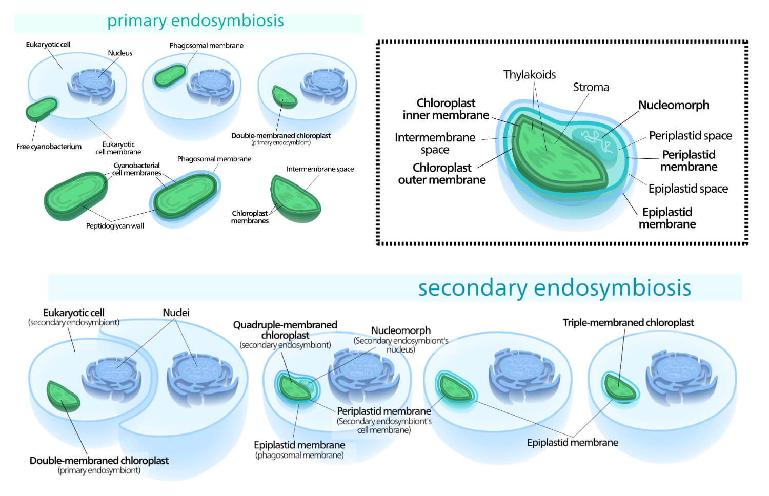
Evidence that Shows the Existence of Endosymbiosis in Eukaryotic Cells
The evolution of cells to eukaryotic is believed due to the involvement of endosymbiosis of organelles such as plastids, and mitochondria (that were initially prokaryotes) which later on formed the complex eukaryotic cells.
- The cell organelles such as chloroplast, and mitochondria can divide binary fission which might have been separate at first and later symbiotically entered the eukaryotic cell.
- The size of bacterial and those cell organelles is similar.
- The presence of an extra outer membrane may be due to the vesicular transport of these cells into the eukaryotic cell.
- Along with the 80s ribosomes, there is also the presence of the 70s ribosomes which are common in prokaryotes.
- As in the prokaryotes, mitochondria, and chloroplast also have circular and naked DNA.
- The cell organelles also show susceptibility toward some antibiotics such as Chloramphenicol which is the characteristic of bacterial cells.
Endosymbiosis Significances
- The endosymbiont gets a favorable environment for its survival.
- The hosts consume different nutrients which are required by the endosymbiont for their growth, multiplication, and survival.
- The hosts also get some benefits such as some nutrients and anti-pathogenic chemicals from the endosymbiont. For eg. In the case of E. coli , it releases chemicals called colicin which can harm other pathogens entering the human body.
- Endosymbiosis process is also beneficial in the evolution of cells, where one cell moved inside the other and gradually developed into a complex cellular structure. Different forms of life or organisms that currently exist are because of endosymbiosis.
Some Related Terms
Ectosymbiosis: It is the phenomenon in which one organism on the surface or skin or outer body part of another organism and they both have a mutually beneficial relationship. The organisms that show ectosymbiosis are called ectosymbionts.
Protocoopeartion: It is similar to symbiosis but not obligatory.
Commensalism: It is a type of relationship in which one of the members is benefitted while the other is neither benefitted nor harmed. The organisms that show commensalism are called commensals.
Parasitism: It is a type of relationship in which one organism is benefitted while another is harmed due to the presence of that member called a parasite.
- Nowack, Eva C M, and Michael Melkonian. “Endosymbiotic associations within protists.” Philosophical Transactions of the Royal Society of London. Series B, Biological sciences vol. 365,1541 (2010): 699-712.
- Moran N.A., and Chong R.A. (2018) Endosymbiosis. Evolutionary Biology. Doi: 10.1093/OBO/9780199941728-0114
- Martin W.F., Garg S., and Zimorski V. (2015) Endosymbiotic Theories of Eukaryote Origin. Phil. Trans. R. Soc. B 370: 20140330
- Lynn M. (2011), Symbiogenesis. A New Principle of Evolution Rediscovery. Palaentological Journal. 44 (12): 1525-1539.
About Author
Bikash Dwivedi
1 thought on “Endosymbiosis- Definition, 5 Examples, Theory, Significances”
It’s good
Leave a Comment Cancel reply
Save my name, email, and website in this browser for the next time I comment.
- Skip to content
- Skip to secondary menu
- Skip to primary sidebar
- Skip to footer
Health and Medical Blog
Endosymbiotic Theory of the Origin of Eukaryotic Cells
Endosymbiotic theory, which is often referred to as “symbiogenesis,” is an evolutionary theory that attempts to explain the origin of eukaryotic cells. It is a hypothesis which essentially postulates that prokaryotes were what gave rise to the first eukaryotic cells and, if true, would rank amongst the most important evolutionary events in our history.
Eukaryotic cell organelles include mitochondria that is found in animals or fungi and the chloroplasts that are found in plants. Mitochondria are one of several different organelles that are found in the cells of every single eukaryotic cell.
Thanks to sequencing technologies, symbiogenesis has grown in prominence as a theory over the last several decades. There are several competing theories, including non-evolutionary theories, to explain the origin of eukaryotic cells, and the science behind the theory is still in its beginning stages, but it does help to explain the symbiotic relationships that two different species are dependent upon for their mutual survival.
The Details Behind the Origins of Eukaryotic Cells
In the Endosymbiotic theory, the idea is that a eukaryotic mitochondrion evolved from an autotrophic bacterium that had been engulfed by the eukaryotic cell. This cell was able to arise when an anaerobic prokaryote lost its cell wall because it was unable to use oxygen for energy. The flexible membrane underneath began to grow and then fold in on itself, which led to the creation of a nucleus and additional internal membranes.
In other words, the eukaryote cell would eat the prokaryote, but would not actually digest it. It would instead keep the bacterium in a symbiotic relationship so that the two co-exist together. The symbiont would then begin to lose some of its genetic material as it forms into a mitochondrion.
This process would then continue because the eukaryote and mitochondria are still existing in a symbiotic relationship. The theory holds that the eukaryote and mitochondria symbiont would eat an autotrophic eukaryote cell, but again not actually digest it. As before, the new cell would also be kept as a symbiont. The secondary symbiont would also lose some of its genetic material during this process and this would be the foundation of the creation of a chloroplast.
It is believed that this process would have occurred in the earlier days of our planet’s history. It could be as much as 2.5 billion years ago. By having the prokaryote utilizing cellular respiration to convert organic molecules into energy, the cells would be too small to be digested by a eukaryotic cell, but would instead live inside the hose cell, contributing to the host’s eventual evolution.
When Was Endosymbiotic Theory First Proposed?
Endosymbiotic theory was first proposed by Konstantin Mereschkowski in 1910. He worked as a botanist and through his work, found the ideas described above to be plausible. It would be more than 50 years before the microbiological evidence discovered by Lynn Margulis in 1967 would help to substantiate the theory.
Mereschkowski primarily researched lichens and had previously published some of the initial fundamentals of symbiogenesis in a 1905 paper that explored the origins and nature of chromatophores in the plant kingdom.
Lichens were his primary point of fascination in the development of this theory because he found that there was a symbiotic relationship between algae and fungi. With over 2,000 specimens collected, it is still in the possession of Kazan University where much of his work takes place.
Evidence from biochemical and molecular sources suggests that mitochondria were developed from proteobacteria and chloroplasts came from cyanobacteria, which would eventually help to form the backbone of life on our planet as we know it.
How Endosymbiotic Theory Influences Evolution
When many discuss the theory of evolution, it is usually the theory that was proposed by Charles Darwin that is looked at. Endosymbiotic theory is more of a rebuttal of Darwinian evolution because it does not rely on the idea that natural selection can explain the full branches of biological novelty. Symbiogenesis offers an alternative where acquisition and inheritance of microbes is the primary life development factor.
The evidence to support Endosymbiotic theory is a list that is quite lengthy.
- Mitochondria and plastids are formed through a process that is similar to binary fission, which is the form of cell division that bacteria use.
- When mitochondria or chloroplasts are removed from a cell, then the cell loses the ability to create new ones.
- Porins are found in the outer membranes of mitochondria, chloroplasts, and bacterial cells.
- Certain mitochondria and plastids contain a single, circular DNA molecule that is similar to bacterial DNA is size and structure.
- Mitochondria and plastids have small genomes when compared to bacteria. Neither organelle is capable of surviving outside the cell, which supports the idea of increased dependence as offered through Endosymbiotic theory.
- Ribosomes found in mitochondria and plastids are more like those found in bacteria than in eukaryotes.
- Some species of lice have multiple chromosomes in the mitochondrion. This, along with other genetic evidence, suggests that mitochondria have multiple ancestors that were acquired by symbiogenesis on several different occasions instead of just one single incident.
When looking at Endosymbiotic theory through modern science, it appears that there were extensive mergers and rearrangements of genetic material in several of the original mitochondrial chromosomes. In looking at lice at this level, it shows that the symbiotic relationships in the ancient world could have formed to work together in a way that could create the building blocks of life as we know it on our planet today.
How Endosymbiotic Theory Affects Creationism
In many circles, the idea that evolution and creationism could work together is seen as absurd. They are often treated as two conflicting theories. Yet the processes described in Endosymbiotic theory correspond with the creation processes that are described within the first chapter of the book of Genesis.
This means there must be a separation between “Old World” creationists and “New World” creationists when it comes to looking at actual creation theory. New World creationists view the book of Genesis literally, with creation taking 6 literal days. From an Old World perspective, the “days” mentioned in Genesis could be an indeterminate amount of time.
This means the process of Endosymbiotic theory for the origin of eukaryotic cells could help to explain how God created the world just as they would help to explain how the world naturally evolved on its how without supernatural influence.
Why You Need to Get to Know Angomonas Deanei
Angomonas denai is a protozoan. This parasite is found in the GI tract of insects. In a closer inspection of this parasite, it has been found that it is a hose to symbiotic bacteria. The symbiont relationship is so extensive, in fact, that the two have formed a permanent relationship. Neither can survive on its own without the other.
This relationship serves as a model for evidence of endosymbiotic theory in practice today in nature. It was first discovered in 1973 and its cell membranes exhibit unusual features that include membrane lipids that are mostly present in the symbiotic prokaryotes of eukaryotic cells.
The One Issue with the Endosymbiotic Theory
When we discuss evolutionary theories or creationism, the goal is often an attempt to explain the origin of the universe. The fact is that no one understands what the universe was like right before or right after matter was created. At some point, primordial energy, space, and time had to arise somehow. The Endosymbiotic theory does not make an attempt to explain the start of the universe.
It instead attempts to explain the start of how life as we know it was able to form.
Theories are mathematical models that allow us to be able to make a prediction about the world and how it behaves. At the quantum level, which is sub-atomic, we can make predictions about behavior in terms of distance, but not about how the universe itself formed.
This is why it is important to understand what the purpose of a theory happens to be. For some, the creation of the universe is tied to the creation of the planet. For others, these are two separate events that require two separate theories to create a plausible explanation.
It could be that life on our planet is unique because of the factors explained through Endosymbiotic theory. It could also mean that the origin of eukaryotic cells is very common throughout the universe and that life is evolving in unique ways on many different planets that have yet to be discovered.
Our universe may be entirely unique. It may just be one of many universes that exist on a scale that goes beyond anything that exists in our imagination. Some may feel that the universe was made for us, while others believe that we were made through a natural process. The bottom line is this: we exist. How we exist may be explained by the endosymbiotic theory of the origin of eukaryotic cells or some other theory.
We must keep asking questions. Only when we continue to seek with an open mind will we be able to find our answers.
- 13 ANC Nails Pros and Cons
- 15 Artificial Sphincter Pros and Cons
- 14 Hysterectomy for Fibroids Pros and Cons
- 15 Monovision Lasik Pros and Cons
- 12 Pros and Cons of the Da Vinci Robotic Surgery
- 14 Peritoneal Dialysis Pros and Cons
- 14 Pros and Cons of the Cataract Surgery Multifocal Lens
- 19 Dermaplaning Pros and Cons
- 15 Mirena IUD Pros and Cons
- 11 Pros and Cons of Monovision Cataract Surgery
- Calories Burned
- Cancer Articles and Infographics
- Definitions and Examples of Theory
- Definitions for Kids
- Dental Articles and Infographics
- Elder Care Articles and Infographics
- Environmental
- Health Research Funding
- Healthcare Articles and Infographics
- ICD 9 Codes
- Major Accomplishments
- Medical Articles and Infographics
- Nutrition Articles and Infographics
- Pharmaceutical Articles and Infographics
- Psychological Articles and Infographics
- Skin Articles and Infographics
- Surgery Articles and Infographics
- Theories and Models
- Uncategorized
- Videos on How to Get Research Funding
Biology Simple
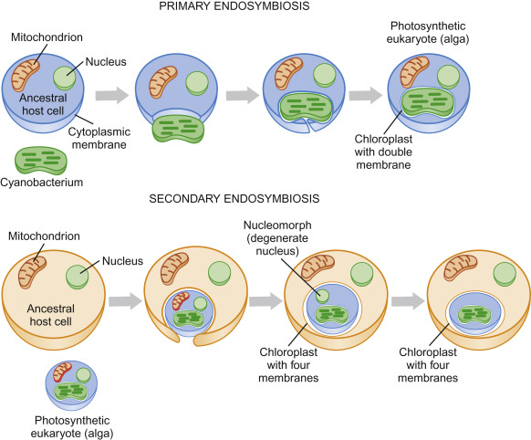
Endosymbiosis
Endosymbiosis is a process in which one organism lives inside another and both benefit. It is an important concept in evolutionary biology and has led to the development of many complex organisms.
Endosymbiosis is a crucial mechanism for the evolution of eukaryotic cells, involving the engulfment of one prokaryotic cell by another. This process has given rise to the mitochondria and chloroplasts, which are essential components of eukaryotic cells. The establishment of these symbiotic relationships has significantly contributed to the diversity and complexity of life on Earth.
Understanding endosymbiosis provides valuable insight into the interconnectedness of organisms and the evolutionary processes that have shaped the natural world. As such, it is a fundamental concept in biology and continues to be a focus of scientific research and study.
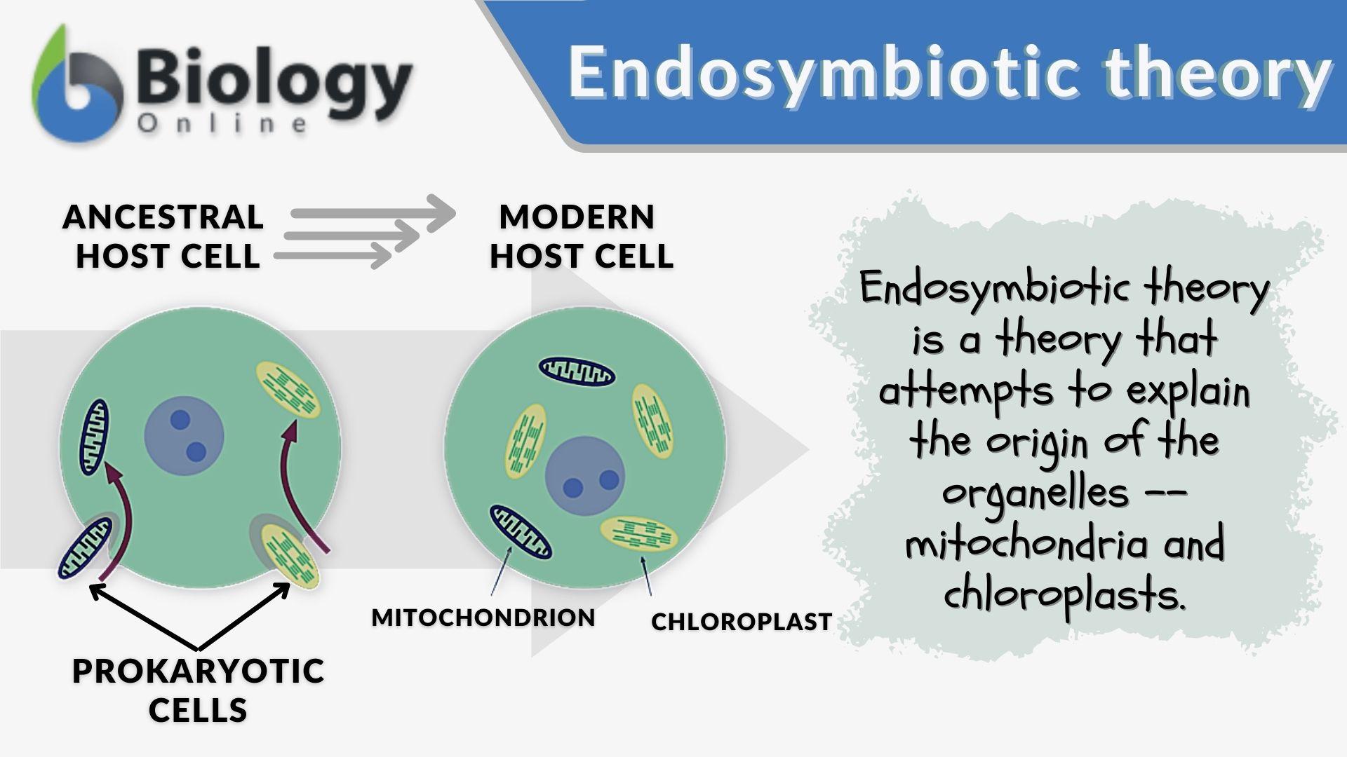
Credit: www.biologyonline.com
The Origin Of Endosymbiotic Theory
Endosymbiosis, the process by which a cell engulfs another cell, leading to a mutually beneficial relationship, has been a fascinating subject of study in the field of evolutionary biology. The origin of endosymbiotic theory traces back to the groundbreaking work of several scientists who made significant contributions to our understanding of this phenomenon. Let’s delve into the early observations and the subsequent development of the theory that has revolutionized our understanding of cellular evolution.
Early Observations
In the late 19th century, the German botanist Andreas Schimper and later, the Russian biologist Konstantin Mereschkowski, independently observed similarities between chloroplasts and mitochondria within eukaryotic cells and free-living prokaryotic organisms. These observations laid the groundwork for the endosymbiotic theory , proposing that these organelles were once free-living prokaryotes that were engulfed by ancestral eukaryotic cells.
Development Of The Theory
The endosymbiotic theory gained further support through the work of Lynn Margulis in the 1960s and 1970s. Margulis provided substantial evidence to support the idea that mitochondria and chloroplasts were once free-living bacteria , and through a process of symbiosis, became integrated into eukaryotic cells. Her groundbreaking research and publications solidified the endosymbiotic theory as a key framework for understanding the origins of organelles within eukaryotic cells, fundamentally altering our perception of the intricate relationships between different cellular entities.

Examples Of Endosymbiosis In Nature
Endosymbiosis is a fascinating phenomenon where one organism lives inside another, forming a mutually beneficial relationship. This incredible process is found abundantly in nature and has played a crucial role in the evolution of various species. Let’s explore some prominent examples of endosymbiosis:
Mitochondria
Mitochondria are small organelles found in the cells of most living creatures. They are often referred to as the “powerhouses” of the cell, as they are responsible for generating energy to fuel cellular processes. Interestingly, mitochondria were not always a part of our cells. At some point in ancient evolutionary history, they were independent organisms themselves.
It is theorized that a eukaryotic host cell engulfed a free-living aerobic bacterium, giving rise to the mitochondria we know today. Over time, an endosymbiotic relationship formed between the host cell and the mitochondria. The host provided the necessary environment and protection for the mitochondria, while the mitochondria produced energy for the cell through a process called cellular respiration.
Chloroplasts
Similar to mitochondria, chloroplasts are organelles found in plants and algae. They play a vital role in photosynthesis, the process by which plants convert sunlight into energy. Like mitochondria, chloroplasts were once independent organisms that formed an endosymbiotic relationship with eukaryotic cells.
Through endosymbiosis, a eukaryotic cell engulfed a photosynthetic cyanobacterium, giving rise to chloroplasts. Over time, the cell and the chloroplasts established a mutually beneficial relationship. The cell provided the necessary environment and protection for the chloroplasts, while the chloroplasts produced energy-rich compounds through photosynthesis, enabling the cell to thrive.
Other Symbiotic Relationships
Endosymbiosis extends beyond mitochondria and chloroplasts. Many other symbiotic relationships can be found in nature, each demonstrating the power of collaboration and interdependence between organisms.
For example, in lichens, a mutualistic association between fungi and photosynthetic algae or cyanobacteria allows them to thrive in diverse environments. The fungi provide a protective structure and absorb water and nutrients, while the algae or cyanobacteria contribute to the partnership by producing energy through photosynthesis.
Another fascinating example is the relationships between some marine animals and bioluminescent bacteria. These bacteria live symbiotically within the host, providing the ability to produce light, which can offer camouflage, attract prey, or even aid in communication among individuals of a species.
Symbiotic relationships, including endosymbiosis, are essential for the evolution and survival of species. From life’s earliest stages on Earth to the intricate ecosystems we see today, these examples of endosymbiosis demonstrate the extraordinary interconnectedness of life forms and the remarkable possibilities that arise from cooperation.
Mechanisms Of Endosymbiosis
Endosymbiosis is a fascinating phenomenon in which one organism lives inside another, forming a mutually beneficial relationship. This intriguing process relies on various mechanisms that enable the integration of different genetic materials, leading to the cooperative coexistence between the host and the endosymbiont. Understanding the mechanisms of endosymbiosis can shed light on the intricate relationships between organisms and offer insights into the evolution of complex life forms.
Process Of Endosymbiosis
The process of endosymbiosis involves several steps that culminate in the establishment of a stable association between the host and endosymbiont. Let’s take a closer look at each of these steps:
- Initial Contact: The first crucial step in endosymbiosis occurs when the host and the endosymbiont come into contact. This interaction can be facilitated through various means, such as physical contact, mutual attraction, or chemical signaling.
- Invasion: Once contact is established, the endosymbiont must find its way to the host’s interior. This can occur through active penetration or by being engulfed by the host’s cell.
- Host Recognition: Host recognition is a pivotal stage, as the host must distinguish between beneficial and harmful organisms. This recognition process often involves molecular signaling that allows the host to identify compatible endosymbionts.
- Establishment of Residence: Upon successful recognition, the endosymbiont establishes residence within the host’s cell or tissue. This integration typically involves physical and biochemical changes in both the host and the endosymbiont.
- Mutual Adaptation: As the endosymbiont resides within the host, both organisms undergo a process of mutual adaptation. Genetic changes occur in both the host and the endosymbiont, leading to a coordinated interaction that benefits both parties.
Genetic Integration
The genetic integration between the host and the endosymbiont is a key aspect of the endosymbiotic relationship. This integration may involve the transfer of genetic material from one organism to another, resulting in genetic changes that contribute to their mutual interaction and cooperation.
One mechanism of genetic integration in endosymbiosis is through horizontal gene transfer. Horizontal gene transfer is the transfer of genetic material between different organisms that are not direct offspring. This horizontal transfer of genes allows for the acquisition of new traits and adaptations, leading to the evolution of more complex organisms.
Additionally, endosymbiosis can also result in gene loss or reduction, where redundant genes from either the host or the endosymbiont are no longer necessary because of their mutual cooperation. This reduction in gene content streamlines the genomes of both organisms, enhancing their overall efficiency and coordination.
In conclusion, the mechanisms of endosymbiosis involve a series of steps, including initial contact, invasion, host recognition, establishment of residence, and mutual adaptation. These mechanisms, coupled with genetic integration, shape the intricate relationships between the host and endosymbiont, leading to their successful coexistence. Understanding these mechanisms provides remarkable insights into the complex evolution of life on our planet.

Credit: microbenotes.com
Evolutionary Implications
Endosymbiosis, the process by which a symbiotic relationship is established between two distinct organisms, has profound evolutionary implications. This process is believed to have played a pivotal role in shaping the cellular evolution and biodiversity of life on Earth.
Impact On Cellular Evolution
The incorporation of endosymbiotic bacteria into early eukaryotic cells is a significant event in cellular evolution. This fusion provided eukaryotic cells with novel metabolic capabilities, allowing for the development of more complex cellular structures and functions. It contributed to the emergence of mitochondria and chloroplasts, critical organelles that are vital for cellular respiration and photosynthesis, respectively.
Influence On Biodiversity
Endosymbiosis has had a profound influence on the diversity of life forms on our planet. The acquisition of these symbiotic relationships has allowed organisms to adapt to diverse ecological niches, contributing to the proliferation of diverse species. Additionally , endosymbiosis has facilitated the evolution of mutualistic relationships, enabling organisms to thrive in various environments and coevolve with their host organisms.
Controversies And Debates
Endosymbiosis theory has sparked controversies and debates in the scientific community.
Challenges To The Theory
Some scientists question the feasibility of endosymbiosis due to lack of experimental evidence .
- Challenges arise from the complexity of cellular evolution.
- Doubts stem from the difficulty in replicating endosymbiotic events.
Alternative Explanations
Researchers propose alternative theories to explain organelle origins.
- One suggestion is the serial endosymbiosis theory that posits a staged evolution .
- Another view points to horizontal gene transfer as a potential driver of cell evolution.
Applications Of Endosymbiotic Theory
The applications of endosymbiotic theory, which revolves around the concept of endosymbiosis, are diverse and significant. It explains how certain organelles in eukaryotic cells, such as mitochondria and chloroplasts, were once independent prokaryotic organisms that formed a symbiotic relationship with host cells.
This theory has far-reaching implications in fields like evolutionary biology, genetics, and biotechnology.
Biotechnological Advances
Medical relevance, future prospects.
The future prospects of endosymbiosis present exciting opportunities for further research and technological innovations. Understanding the mechanisms and implications of endosymbiosis can lead to significant advancements in various scientific fields. From exploring new research directions to technological innovations, the future of endosymbiosis offers a wealth of potential for advancements.
Research Directions
Advancing research into endosymbiosis will continue to uncover new insights into the intricate relationship between host and symbiont. Future studies may explore the potential for harnessing endosymbiotic relationships for sustainable agricultural practices and environmental conservation. Moreover, investigating the role of endosymbiosis in human health and disease can open new avenues for medical interventions and therapeutics.
Technological Innovations
The future of endosymbiosis holds promise for technological innovations that can revolutionize various fields. Advancements in imaging technologies, such as super-resolution microscopy, can provide unprecedented visualization of endosymbiotic interactions at the cellular and molecular levels. Furthermore, advancements in genetic engineering and synthetic biology may allow for the manipulation and optimization of endosymbiotic relationships for beneficial outcomes in diverse applications.

Credit: www.sciencedirect.com
Frequently Asked Questions For Endosymbiosis
What is endosymbiosis in biology.
Endosymbiosis is a process where one organism lives inside another, forming a mutually beneficial relationship.
How Does Endosymbiosis Contribute To Evolution?
Endosymbiosis is believed to have played a significant role in the evolution of eukaryotic cells.
Can Endosymbiosis Lead To New Organisms?
Yes, endosymbiosis can result in the creation of novel organisms with unique characteristics and adaptations.
Endosymbiosis has revolutionized our understanding of the origins and diversity of life on Earth. This intricate process between different organisms has shaped the intricate web of life that exists today. By delving into the fascinating world of endosymbiosis, we gain insights into the symbiotic relationships that have driven the evolution of countless species.
From mitochondria to chloroplasts, these cellular mergers have paved the way for the complexity and diversity of life we see around us. Exploring the wonders of endosymbiosis not only expands our knowledge of biology but also highlights the interconnectedness of all living organisms on our planet.
Embracing the concept of symbiosis allows us to appreciate the delicate balance and cooperation that underpins nature’s remarkable tapestry.
Similar Posts
Liliopsida is a class of flowering plants, also known as monocotyledons, with parallel-veined leaves and flower parts in multiples of three. Liliopsida, commonly referred to as monocotyledons, is a class of flowering plants distinguished by their parallel-veined leaves and flower parts in multiples of three. These plants encompass a diverse range of species, including grasses,…
Ceruloplasmin
Ceruloplasmin is a protein that helps transport copper in the blood, playing a crucial role in copper metabolism. It also has antioxidant properties, protecting cells from damage. Ceruloplasmin serves as a essential enzyme in various biological processes, and its levels can indicate certain health conditions. Just like other proteins, ceruloplasmin is synthesized in the liver…
Lacrimation
Lacrimation is the production of tears to lubricate the eyes and keep them moist. It is a natural part of eye health and helps to protect the eyes from irritants and maintain clear vision. Lacrimation, commonly known as tearing, is a natural process that keeps the eyes moist and healthy. Tears are produced to clean…
Chemical Biology
Chemical Biology studies the chemical processes within biological systems. It explores interactions between molecules in living organisms. Chemical Biology combines chemistry and biology to understand how molecules and cells interact and function in living organisms. This interdisciplinary field aims to develop new tools, techniques, and medicines by investigating the chemical basis of biological processes. Researchers…
Dark Respiration
Dark respiration refers to the process of cellular respiration that occurs in the absence of light, during which organisms break down glucose to produce energy. We will explore the concept of dark respiration and its significance in various organisms. Dark respiration, also known as cellular respiration in the absence of light, is a crucial metabolic…
Bryophytes are non-vascular land plants that include mosses, liverworts, and hornworts and are an important component of damp habitats. These plants have a thallus-like body attached to the substrate by rhizoids and lack true vegetative structures. They reproduce through gametes and have a haploid main plant body called a gametophyte. Dioecious bryophytes have separate male…
Leave a Reply Cancel reply
Your email address will not be published. Required fields are marked *
Save my name, email, and website in this browser for the next time I comment.

Kathleen A. Marrs, Indiana University-Purdue University at Indianapolis

This resource is no longer officially part of our collection This resource has been removed from our collection, likely because the original resource is no longer available. If you have further information about the link (e.g. a new location where the information can be found) please let us know. You may be able to find previous versions at the Internet Archive.
- Microbial Life:Resources for K-12 Teachers and Students:The Microbes Within: Resources

- About this Site
- Accessibility
Citing and Terms of Use
Material on this page is offered under a Creative Commons license unless otherwise noted below.
Show terms of use for text on this page »
Show terms of use for media on this page »
- Short URL: http://serc.carleton.edu/resources/38722.html What's This?
The Endosymbiotic Hypothesis Just another WordPress.com site
Evidence for the endosymbiotic hypothesis.
- History: The Formation of the Endosymbiotic Hypothesis
- Primary versus Secondary Endosymbiosis
Posted by endosymbiotichypothesis.
Similarities Between Bacteria and Semiautonomous Organelles
Since the symbiotic hypothesis states that mitochondria and chloroplasts arose from bacteria entering a eukaryotic cell to form a symbiotic relationship, similarities between bacteria and these semiautonomous organelles show strong evidence that this hypothesis is correct.
Mitochondria share very similar characteristics with purple-aerobic bacteria. They both use oxygen in the production of ATP, and they both do this by using the Kreb’s Cycle and oxidative phosphorylation. (mitochondria on the left and purple aerobic bacteria on the right)

Chloroplasts are very similar to photosynthetic bacteria in that they both have very similar chlorophyll that harness light energy to convert into chemical energy. (Chloroplast on the left and photosynthetic bacteria on the right)

Although there are many similarities between mitochondria and purple aerobic bacteria and chloroplasts and photosynthetic bacteria, they appear to be slight and to have arisen via evolution.
Size of mitochondria and chloroplasts in comparison to bacteria is another simple observation that supports the endosymbiotic hypothesis. Mitochondria, chloroplasts, and prokaryotes (bacteria) range from about one to ten microns in size. (1 micron=1X10 -6 Meters) This seems very basic, but if there was a large difference in sizes between these three components, the hypothesis would appear to be false.
- DNA, RNA, Ribosomes and Protein Synthesis
The first piece of evidence that needed to be found to support the endosymbiotic hypothesis was whether or not mitochondria and chloroplasts have their own DNA and if this DNA is similar to bacterial DNA. This was later proven to be true for DNA, RNA, ribosomes, chlorophyll (for chloroplasts), and protein synthesis. This provided the first substantial evidence for the endosymbiotic hypothesis. It was also determined that mitochondria and chloroplasts divide independently of the cell they live in.
Mitochondria having their own DNA and dividing independently of the cell is what ultimately results in only mitochondrial DNA being inherited by one’s mother since only an egg cell has DNA while a sperm cell does not. (This relationship also further proves that the discovered characteristics of mitochondria are true.)
This level of independence among semiautonomous organelles shows that they are not very related to the nucleus or other organelles of a eukaryotic cell. Since they are not related, it appears to be even more probable that mitochondria and chloroplasts were originally bacteria that entered the eukaryotic cell via endocytosis to form a symbiotic relationship.
Evolutionary Drive
Scientists (particularly Lynn Margulis) then began to think that if mitochondria and chloroplasts were truly bacteria that were taken into eukaryotic cells via endocytosis, then there must be a historical drive to promote this symbiotic relationship. About 3.8 billion years ago, there were only anaerobic bacteria in existence because Earth’s atmosphere did not contain any oxygen. The first photosynthetic bacteria arose around 3.2 billion years ago and began producing large quantities of oxygen as a byproduct of photosynthesis. Oxygen is very toxic to cells, and as a result, these anaerobic, photosynthetic bacteria became less effective at surviving in their environment. At this point, some of the anaerobic bacteria evolved into aerobic bacteria. Aerobic bacteria are much better suited to this oxygen containing environment and they even use oxygen in the process of making ATP (a molecule that stores a great amount of easily accessible energy). One important factor that both of these bacteria lacked was the ability to ingest large quantities of nutrients from the surrounding environment via phagocytosis. About 1.5 billion years ago, the first nucleated cell (the eukaryote) was arose through evolution, and this cell had the groundbreaking ability to take in large quantities of nutrients via phagocytosis. The fact that bacteria, which are very similar to mitochondria and chloroplasts, existed before the eukaryotic cell shows evidence that it was bacteria that was integrated into a eukaryotic cell rather than eukaryotes being entirely separate in evolutionary history. This timeline also gives evidence as to why a symbiotic relationship would be beneficial.
The photosynthetic and aerobic bacteria were naturally driven to enter into this relationship because the eukaryotic cell supplies both protection and nutrients, and the bacteria supply ways for eukaryotes to harness more energy than they previously could using only glycolysis.

This (above) is the second stage of the glycolysis process (the only stage that actually produces the ATP), and as you can see it only produces a total of 4 ATP (2 net ATP). When this process is combined with the Krebs cycle and oxidative phosphorylation (which requires mitochondria), the net amount of ATP produced is 36-38 molecules.
By eukaryotic cells engulfing photosynthetic bacteria, they could then create glucose molecules that could then be used to go through the catabolic processes in the mitochondria, and hence, the eukaryotic cell harnesses even more energy than it would on its own. Having so much energy to drive cellular processes makes this new eukaryotic cell more fit for survival.
Double Phospholipid Bilayer
A fairly simple piece of evidence for the endosymbiotic hypothesis is the fact that both mitochondria and chloroplasts have double phospholipid bilayers. This appears to have arisen by mitochondria and chloroplasts entering eukaryotic cells via endocytosis. Both purple, aerobic bacteria (similar to mitochondria) and photosynthetic bacteria (similar to chloroplasts) only have one phospholipid bilayer, but when they enter another cell via endocytosis, they are bound by a vesicle which forms the second layer of their double phospholipid bilayer.
This video shows the process of endocytosis of aerobic bacteria and photosynthetic bacteria very well.
Share this:
Leave a comment cancel reply, latest posts.
- The Endosymbiotic Hypothesis
- Documentation
- Suggest Ideas
- Support Forum
- WordPress Blog
- WordPress Planet
Blog at WordPress.com.

- Already have a WordPress.com account? Log in now.
- Subscribe Subscribed
- Copy shortlink
- Report this content
- View post in Reader
- Manage subscriptions
- Collapse this bar
Mitochondria: fundamental characteristics, challenges, and impact on aging
- Review Article
- Published: 28 August 2024
Cite this article

- Runyu Liang 1 ,
- Luwen Zhu 2 ,
- Yongyin Huang 1 ,
- Jia Chen 1 &
- Qiang Tang 2
As one of the most vital organelles within biological cells, mitochondria hold an irreplaceable status and play crucial roles in various diseases. Research and therapies targeting mitochondria have achieved significant progress in numerous conditions. Throughout an organism’s lifespan, mitochondrial dynamics persist continuously, and due to their inherent characteristics and various external factors, mitochondria are highly susceptible to damage. This susceptibility is particularly evident during aging, where the decline in biological function is closely intertwined with mitochondrial dysfunction. Despite being an ancient and enigmatic organelle, much remains unknown about mitochondria. Here, we will explore the past and present knowledge of mitochondria, providing a comprehensive review of their intrinsic properties and interactions with nuclear DNA, as well as the challenges and impacts they face during the aging process.
This is a preview of subscription content, log in via an institution to check access.
Access this article
Subscribe and save.
- Get 10 units per month
- Download Article/Chapter or eBook
- 1 Unit = 1 Article or 1 Chapter
- Cancel anytime
Price includes VAT (Russian Federation)
Instant access to the full article PDF.
Rent this article via DeepDyve
Institutional subscriptions
Mitochondrial Function in Aging
Mitophagy plays a central role in mitochondrial ageing.

Mitophagy, Diseases, and Aging
Data availability.
No datasets were generated or analysed during the current study.
Akbari M, Nilsen HL, Montaldo NP (2022) Dynamic features of human mitochondrial DNA maintenance and transcription. Front Cell Dev Biol 10:984245. https://doi.org/10.3389/fcell.2022.984245
Article PubMed PubMed Central Google Scholar
Amorim JA, Coppotelli G, Rolo AP et al (2022) Mitochondrial and metabolic dysfunction in ageing and age-related diseases. Nat Rev Endocrinol 18:243–258. https://doi.org/10.1038/s41574-021-00626-7
Anderson NS, Haynes CM (2020) Folding the mitochondrial UPR into the integrated stress response. Trends Cell Biol 30:428–439. https://doi.org/10.1016/j.tcb.2020.03.001
Article CAS PubMed PubMed Central Google Scholar
Angelini C, Bello L, Spinazzi M, Ferrati C (2009) Mitochondrial disorders of the nuclear genome. Acta Myol Myopathies Cardiomyopathies Off J Mediterr Soc Myol 28:16–23
CAS Google Scholar
Behl T, Makkar R, Anwer MK et al (2023) Mitochondrial dysfunction: a cellular and molecular hub in pathology of metabolic diseases and infection. J Clin Med 12:2882. https://doi.org/10.3390/jcm12082882
Bergamini C, Bonora E, Moruzzi N (2023) Editorial: mitochondrial bioenergetics impairments in genetic and metabolic diseases. Front Physiol 14:1228926. https://doi.org/10.3389/fphys.2023.1228926
Betancourt AM, King AL, Fetterman JL et al (2014) Mitochondrial-nuclear genome interactions in non-alcoholic fatty liver disease in mice. Biochem J 461:223–232. https://doi.org/10.1042/BJ20131433
Article CAS PubMed Google Scholar
Boguszewska K, Szewczuk M, Kaźmierczak-Barańska J, Karwowski BT (2020) The similarities between human mitochondria and bacteria in the context of structure, genome, and base excision repair system. Mol Basel Switz 25:2857. https://doi.org/10.3390/molecules25122857
Article CAS Google Scholar
Chang C-W, Xu X, Li M et al (2020) Pathogenic mutations reveal a role of RECQ4 in mitochondrial RNA:DNA hybrid formation and resolution. Sci Rep 10:17033. https://doi.org/10.1038/s41598-020-74095-9
Chauhan A, Vera J, Wolkenhauer O (2014) The systems biology of mitochondrial fission and fusion and implications for disease and aging. Biogerontology 15:1–12. https://doi.org/10.1007/s10522-013-9474-z
Chinnery PF, Prudent J (2019) De-fusing mitochondria defuses the mtDNA time-bomb. Cell Res 29:781–782. https://doi.org/10.1038/s41422-019-0206-z
Covarrubias AJ, Perrone R, Grozio A, Verdin E (2021) NAD + metabolism and its roles in cellular processes during ageing. Nat Rev Mol Cell Biol 22:119–141. https://doi.org/10.1038/s41580-020-00313-x
Dai C-Y, Ng CC, Hung GCC et al (2023) ATFS-1 counteracts mitochondrial DNA damage by promoting repair over transcription. Nat Cell Biol 25:1111–1120. https://doi.org/10.1038/s41556-023-01192-y
Das BB, Ghosh A, Bhattacharjee S, Bhattacharyya A (2021) Trapped topoisomerase-DNA covalent complexes in the mitochondria and their role in human diseases. Mitochondrion 60:234–244. https://doi.org/10.1016/j.mito.2021.08.017
Devin A, Rigoulet M (2007) Mechanisms of mitochondrial response to variations in energy demand in eukaryotic cells. Am J Physiol Cell Physiol 292:C52–58. https://doi.org/10.1152/ajpcell.00208.2006
DiMauro S, Schon EA (2003) Mitochondrial respiratory-chain diseases. N Engl J Med 348:2656–2668
Dong L, Neuzil J (2019) Targeting mitochondria as an anticancer strategy. Cancer Commun Lond Engl 39:63. https://doi.org/10.1186/s40880-019-0412-6
Article Google Scholar
Du S-H, Shi J, Yu T-Y et al (2022) Nicotinamide mononucleotide ameliorates acute lung injury by inducing mitonuclear protein imbalance and activating the UPRmt. Exp Biol Med Maywood NJ 247:1264–1276. https://doi.org/10.1177/15353702221094235
Dunham-Snary KJ, Ballinger SW (2015) GENETICS. Mitochondrial-nuclear DNA mismatch matters. Science 349:1449–1450. https://doi.org/10.1126/science.aac5271
Fairbrother-Browne A, Ali AT, Reynolds RH et al (2021) Mitochondrial-nuclear cross-talk in the human brain is modulated by cell type and perturbed in neurodegenerative disease. Commun Biol 4:1262. https://doi.org/10.1038/s42003-021-02792-w
Fang C, Wei X, Wei Y (2016a) Mitochondrial DNA in the regulation of innate immune responses. Protein Cell 7:11–16. https://doi.org/10.1007/s13238-015-0222-9
Fang EF, Scheibye-Knudsen M, Chua KF et al (2016b) Nuclear DNA damage signalling to mitochondria in ageing. Nat Rev Mol Cell Biol 17:308–321. https://doi.org/10.1038/nrm.2016.14
Feric M, Demarest TG, Tian J et al (2021) Self-assembly of multi-component mitochondrial nucleoids via phase separation. EMBO J 40:e107165. https://doi.org/10.15252/embj.2020107165
Fernández-Moreno MA, Vázquez-Fonseca L, Zambrano SP, Garesse R (2021) Mitochondrial DNA: defects, maintenance genes and depletion. Mitochondrial Dis Theory Diagn Ther 2021:69–94
Fernandez-Vizarra E, Zeviani M (2021) Mitochondrial disorders of the OXPHOS system. FEBS Lett 595:1062–1106. https://doi.org/10.1002/1873-3468.13995
Fetterman JL, Zelickson BR, Johnson LW et al (2013) Mitochondrial genetic background modulates bioenergetics and susceptibility to acute cardiac volume overload. Biochem J 455:157–167. https://doi.org/10.1042/BJ20130029
Franchini D-M, Petersen-Mahrt SK (2014) AID and APOBEC deaminases: balancing DNA damage in epigenetics and immunity. Epigenomics 6:427–443. https://doi.org/10.2217/epi.14.35
Gao X, Yu X, Zhang C et al (2022) Telomeres and mitochondrial metabolism: implications for cellular senescence and age-related diseases. Stem Cell Rev Rep 18:2315–2327. https://doi.org/10.1007/s12015-022-10370-8
García-Lepe UO, Bermúdez-Cruz RM (2019) Mitochondrial genome maintenance: damage and repair pathways. DNA Repair- Updat
Garibotti MC, Perry CGR (2023) Strength athletes and mitochondria: it’s about time. J Physiol 601:2753–2754. https://doi.org/10.1113/JP284856
Gattermann N (2004) Mitochondrial DNA mutations in the hematopoietic system. Leukemia 18:18–22. https://doi.org/10.1038/sj.leu.2403209
George A, Aubol BE, Fattet L, Adams JA (2019) Disordered protein interactions for an ordered cellular transition: Cdc2-like kinase 1 is transported to the nucleus via its Ser-Arg protein substrate. J Biol Chem 294:9631–9641. https://doi.org/10.1074/jbc.RA119.008463
Gibellini L, Moro L (2021) Programmed cell death in health and disease. Cells 10:1765. https://doi.org/10.3390/cells10071765
Granat L, Hunt RJ, Bateman JM (2020) Mitochondrial retrograde signalling in neurological disease. Philos Trans R Soc Lond B Biol Sci 375:20190415. https://doi.org/10.1098/rstb.2019.0415
Gray MW (2012) Mitochondrial evolution. Cold Spring Harb Perspect Biol 4:a011403. https://doi.org/10.1101/cshperspect.a011403
Greenberg EF, Vatolin S (2018) Symbiotic origin of aging. Rejuvenation Res 21:225–231. https://doi.org/10.1089/rej.2017.1973
Article PubMed Google Scholar
Gureev AP, Shaforostova EA, Popov VN (2019) Regulation of mitochondrial biogenesis as a way for active longevity: interaction between the Nrf2 and PGC-1α signaling pathways. Front Genet 10:435. https://doi.org/10.3389/fgene.2019.00435
Hämäläinen RH, Landoni JC, Ahlqvist KJ et al (2019) Defects in mtDNA replication challenge nuclear genome stability through nucleotide depletion and provide a unifying mechanism for mouse progerias. Nat Metab 1:958–965. https://doi.org/10.1038/s42255-019-0120-1
Harrington JS, Ryter SW, Plataki M et al (2023) Mitochondria in health, disease, and aging. Physiol Rev 103:2349–2422. https://doi.org/10.1152/physrev.00058.2021
Hazkani-Covo E, Zeller RM, Martin W (2010) Molecular poltergeists: mitochondrial DNA copies (numts) in sequenced nuclear genomes. PLOS Genet 6:e1000834. https://doi.org/10.1371/journal.pgen.1000834
He L, Luo L, Proctor SJ et al (2003) Somatic mitochondrial DNA mutations in adult-onset leukaemia. Leukemia 17:2487–2491. https://doi.org/10.1038/sj.leu.2403146
He Y-H, Chen X-Q, Yan D-J et al (2016) Familial longevity study reveals a significant association of mitochondrial DNA copy number between centenarians and their offspring. Neurobiol Aging. https://doi.org/10.1016/j.neurobiolaging.2016.07.026
Hoang PH, Cornish AJ, Chubb D et al (2020) Impact of mitochondrial DNA mutations in multiple myeloma. Blood Cancer J 10:46. https://doi.org/10.1038/s41408-020-0315-4
Holmbeck MA, Donner JR, Villa-Cuesta E, Rand DM (2015) A Drosophila model for mito-nuclear diseases generated by an incompatible interaction between tRNA and tRNA synthetase. Dis Model Mech 8:843–854. https://doi.org/10.1242/dmm.019323
Hong YS, Battle SL, Shi W et al (2023) Deleterious heteroplasmic mitochondrial mutations are associated with an increased risk of overall and cancer-specific mortality. Nat Commun 14:6113. https://doi.org/10.1038/s41467-023-41785-7
Hopp A-K, Teloni F, Bisceglie L et al (2021) Mitochondrial NAD + controls nuclear ARTD1-induced ADP-ribosylation. Mol Cell 81:340-354e5. https://doi.org/10.1016/j.molcel.2020.12.034
Houtkooper RH, Mouchiroud L, Ryu D et al (2013) Mitonuclear protein imbalance as a conserved longevity mechanism. Nature 497:451–457. https://doi.org/10.1038/nature12188
Hussain M, Mohammed A, Saifi S et al (2023) Hyperubiquitylation of DNA helicase RECQL4 by E3 ligase MITOL prevents mitochondrial entry and potentiates mitophagy. J Biol Chem 299:105087. https://doi.org/10.1016/j.jbc.2023.105087
Irina Z, Elena S, Youri P (2019) Contribution of cytosine desaminases of AID/APOBEC family to carcinogenesis. Biol Commun 64:110–123
Isokallio MA, Stewart JB (2021) High-throughput detection of mtDNA mutations leading to tRNA Processing errors. Methods Mol Biol Clifton NJ 2192:117–132. https://doi.org/10.1007/978-1-0716-0834-0_10
Johnston IG, Williams BP (2016) Evolutionary inference across eukaryotes identifies specific pressures favoring mitochondrial gene retention. Cell Syst 2:101–111. https://doi.org/10.1016/j.cels.2016.01.013
Kaur P, Barnes R, Pan H et al (2021) TIN2 is an architectural protein that facilitates TRF2-mediated trans- and cis-interactions on telomeric DNA. Nucleic Acids Res 49:13000–13018. https://doi.org/10.1093/nar/gkab1142
Kazak L, Reyes A, Holt IJ (2012) Minimizing the damage: repair pathways keep mitochondrial DNA intact. Nat Rev Mol Cell Biol 13:659–671. https://doi.org/10.1038/nrm3439
Kelly G, Kataura T, Panek J et al (2024) Suppressed basal mitophagy drives cellular aging phenotypes that can be reversed by a p62-targeting small molecule. Dev Cell 59:1924–1939 .e7. https://doi.org/10.1016/j.devcel.2024.04.020
Kirby CS, Patel MR (2021) Elevated mitochondrial DNA copy number found in ubiquinone-deficient clk-1 mutants is not rescued by ubiquinone precursor 2-4-dihydroxybenzoate. Mitochondrion 58:38–48. https://doi.org/10.1016/j.mito.2021.02.001
Kolahdouzmohammadi M, Kolahdouz-Mohammadi R, Tabatabaei SA et al (2023) Revisiting the role of autophagy in cardiac differentiation: a comprehensive review of interplay with other signaling pathways. Genes 14:1328. https://doi.org/10.3390/genes14071328
Kulmuni J, Wiley B, Otto SP (2024) On the fast track: hybrids adapt more rapidly than parental populations in a novel environment. Evol Lett 8:128–136. https://doi.org/10.1093/evlett/qrad002
Kumagai H, Miller B, Kim S-J et al (2023) Novel insights into mitochondrial DNA: mitochondrial microproteins and mtDNA variants modulate athletic performance and age-related diseases. Genes 14:286. https://doi.org/10.3390/genes14020286
Kumar R, Harilal S, Parambi DGT et al (2022) The role of mitochondrial genes in neurodegenerative disorders. Curr Neuropharmacol 20:824–835. https://doi.org/10.2174/1570159X19666210908163839
Lauri A, Pompilio G, Capogrossi MC (2014) The mitochondrial genome in aging and senescence. Ageing Res Rev 18:1–15. https://doi.org/10.1016/j.arr.2014.07.001
Lauritzen KH, Olsen MB, Ahmed MS et al (2021) Instability in NAD + metabolism leads to impaired cardiac mitochondrial function and communication. eLife 10:e59828. https://doi.org/10.7554/eLife.59828
Lechuga-Vieco AV, Latorre-Pellicer A, Johnston IG et al (2020) Cell identity and nucleo-mitochondrial genetic context modulate OXPHOS performance and determine somatic heteroplasmy dynamics. Sci Adv 6:eaba5345. https://doi.org/10.1126/sciadv.aba5345
Lechuga-Vieco AV, Justo-Méndez R, Enríquez JA (2021) Not all mitochondrial DNAs are made equal and the nucleus knows it. IUBMB Life 73:511–529. https://doi.org/10.1002/iub.2434
Lee YH, Kuk MU, So MK et al (2023) Targeting mitochondrial oxidative stress as a strategy to treat aging and age-related diseases. Antioxid Basel Switz 12:934. https://doi.org/10.3390/antiox12040934
Leonarduzzi G, Sottero B, Poli G (2010) Targeting tissue oxidative damage by means of cell signaling modulators: the antioxidant concept revisited. Pharmacol Ther 128:336–374. https://doi.org/10.1016/j.pharmthera.2010.08.003
Li TY, Sleiman MB, Li H et al (2021) The transcriptional coactivator CBP/p300 is an evolutionarily conserved node that promotes longevity in response to mitochondrial stress. Nat Aging 1:165–178
Li Y, Li Z, Ren Y et al (2023) Mitochondrial-derived peptides in cardiovascular disease: novel insights and therapeutic opportunities. J Adv Res. https://doi.org/10.1016/j.jare.2023.11.018
Lin Y-H, Lim S-N, Chen C-Y et al (2022) Functional role of mitochondrial DNA in cancer progression. Int J Mol Sci 23:1659. https://doi.org/10.3390/ijms23031659
Lionaki E, Gkikas I, Tavernarakis N (2016) Differential protein distribution between the nucleus and mitochondria: implications in aging. Front Genet. https://doi.org/10.3389/fgene.2016.00162
Loewe L (2006) Quantifying the genomic decay paradox due to Muller’s ratchet in human mitochondrial DNA. Genet Res 87:133–159. https://doi.org/10.1017/S0016672306008123
Marcon F, Bignami M, Karran P (2023) Mutation and aging: news from the pool. Aging 15:4566–4567. https://doi.org/10.18632/aging.204779
Matilainen O, Quirós PM, Auwerx J (2017) Mitochondria and epigenetics - crosstalk in homeostasis and stress. Trends Cell Biol 27:453–463. https://doi.org/10.1016/j.tcb.2017.02.004
Meiklejohn CD, Holmbeck MA, Siddiq MA et al (2013) An incompatibility between a mitochondrial tRNA and its nuclear-encoded tRNA synthetase compromises development and fitness in Drosophila. PLoS Genet 9:e1003238. https://doi.org/10.1371/journal.pgen.1003238
Merkwirth C, Jovaisaite V, Durieux J et al (2016) Two conserved histone demethylases regulate mitochondrial stress-induced longevity. Cell 165:1209–1223. https://doi.org/10.1016/j.cell.2016.04.012
Miller B, Kim S-J, Kumagai H et al (2022) Mitochondria-derived peptides in aging and healthspan. J Clin Invest 132:e158449. https://doi.org/10.1172/JCI158449
Misteli T, Soutoglou E (2009) The emerging role of nuclear architecture in DNA repair and genome maintenance. Nat Rev Mol Cell Biol 10:243–254. https://doi.org/10.1038/nrm2651
Mohrin M, Shin J, Liu Y et al (2015) A mitochondrial UPR-mediated metabolic checkpoint regulates hematopoietic stem cell aging. Science 347:1374–1377. https://doi.org/10.1126/science.aaa2361
Monaghan RM, Whitmarsh AJ (2015) Mitochondrial proteins moonlighting in the nucleus. Trends Biochem Sci 40:728–735. https://doi.org/10.1016/j.tibs.2015.10.003
Moriguchi K, Suzuki T, Ito Y et al (2005) Functional isolation of novel nuclear proteins showing a variety of subnuclear localizations. Plant Cell 17:389–403. https://doi.org/10.1105/tpc.104.028456
Mormone E, Iorio EL, Abate L, Rodolfo C (2023) Sirtuins and redox signaling interplay in neurogenesis, neurodegenerative diseases, and neural cell reprogramming. Front Neurosci 17:1073689. https://doi.org/10.3389/fnins.2023.1073689
Muñoz-Carvajal F, Sanhueza M (2020) The mitochondrial unfolded protein response: a hinge between healthy and pathological aging. Front Aging Neurosci. https://doi.org/10.3389/fnagi.2020.581849
Nargund AM, Pellegrino MW, Fiorese CJ et al (2012) Mitochondrial import efficiency of ATFS-1 regulates mitochondrial UPR activation. Science 337:587–590. https://doi.org/10.1126/science.1223560
Natarajan V, Chawla R, Mah T et al (2020) Mitochondrial dysfunction in age-related metabolic disorders. Proteomics 20:e1800404. https://doi.org/10.1002/pmic.201800404
Needs HI, Protasoni M, Henley JM et al (2021) Interplay between mitochondrial protein import and respiratory complexes assembly in neuronal health and degeneration. Life Basel Switz 11:432. https://doi.org/10.3390/life11050432
Newman LE, Shadel GS (2023) Mitochondrial DNA release in innate immune signaling. Annu Rev Biochem 92:299–332. https://doi.org/10.1146/annurev-biochem-032620-104401
Nguyen NNY, Kim SS, Jo YH (2020) Deregulated mitochondrial DNA in diseases. DNA Cell Biol 39:1385–1400. https://doi.org/10.1089/dna.2019.5220
Pang C-Y, Ma Y-S, Wei Y-U (2008) MtDNA mutations, functional decline and turnover of mitochondria in aging. Front Biosci J Virtual Libr 13:3661–3675. https://doi.org/10.2741/2957
Patel J, den Breems NY, Tuluc M et al (2021) Elevated APOBEC mutational signatures implicate chronic injury in etiology of an aggressive head-and-neck squamous cell carcinoma: a case report. J Med Case Rep 15:252. https://doi.org/10.1186/s13256-021-02685-w
Paull D, Emmanuele V, Weiss KA et al (2013) Nuclear genome transfer in human oocytes eliminates mitochondrial DNA variants. Nature 493:632–637. https://doi.org/10.1038/nature11800
Pichaud N, Bérubé R, Côté G et al (2019) Age dependent dysfunction of mitochondrial and ROS metabolism induced by mitonuclear mismatch. Front Genet 10:130. https://doi.org/10.3389/fgene.2019.00130
Povea-Cabello S, Villanueva-Paz M, Suárez-Rivero JM et al (2020) Advances in mt-tRNA mutation-caused mitochondrial disease modeling: patients’ brain in a dish. Front Genet 11:610764. https://doi.org/10.3389/fgene.2020.610764
Pozzi A, Dowling DK (2021) Small mitochondrial RNAs as mediators of nuclear gene regulation, and potential implications for human health. BioEssays News Rev Mol Cell Dev Biol 43:e2000265. https://doi.org/10.1002/bies.202000265
Pravenec M, Šilhavý J, Mlejnek P et al (2021) Conplastic strains for identification of retrograde effects of mitochondrial dna variation on cardiometabolic traits in the spontaneously hypertensive rat. Physiol Res 70:S471–S484. https://doi.org/10.33549/physiolres.934740
Protasoni M, Serrano M (2023) Targeting mitochondria to control ageing and senescence. Pharmaceutics 15:352. https://doi.org/10.3390/pharmaceutics15020352
Pu W, Gu Z (2023) Incompatibilities between common mtDNA variants in human disease. Proc Natl Acad Sci USA 120:e2217452119. https://doi.org/10.1073/pnas.2217452119
Radzvilavicius A, Layh S, Hall MD et al (2021) Sexually antagonistic evolution of mitochondrial and nuclear linkage. J Evol Biol 34:757–766. https://doi.org/10.1111/jeb.13776
Rattan SIS (2024) Seven knowledge gaps in modern biogerontology. Biogerontology 25:1–8. https://doi.org/10.1007/s10522-023-10089-0
Raval PK, Garg SG, Gould SB (2022) Endosymbiotic selective pressure at the origin of eukaryotic cell biology. eLife 11:e81033. https://doi.org/10.7554/eLife.81033
Reynolds JC, Bwiza CP, Lee C (2020) Mitonuclear genomics and aging. Hum Genet 139:381–399. https://doi.org/10.1007/s00439-020-02119-5
Rudler DL, Hughes LA, Viola HM et al (2021) Fidelity and coordination of mitochondrial protein synthesis in health and disease. J Physiol 599:3449–3462. https://doi.org/10.1113/JP280359
Ryytty S, Modi SR, Naumenko N et al (2022) Varied responses to a high m.3243A > G mutation load and respiratory chain dysfunction in patient-derived cardiomyocytes. Cells 11:2593. https://doi.org/10.3390/cells11162593
San-Millán I (2023) The key role of mitochondrial function in health and disease. Antioxid Basel Switz 12:782. https://doi.org/10.3390/antiox12040782
Sanchez-Contreras M, Kennedy SR (2022) The complicated nature of somatic mtDNA mutations in aging. Front Aging 2:805126. https://doi.org/10.3389/fragi.2021.805126
Sanchez-Contreras M, Sweetwyne MT, Tsantilas KA et al (2023) The multi-tissue landscape of somatic mtDNA mutations indicates tissue-specific accumulation and removal in aging. eLife 12:e83395. https://doi.org/10.7554/eLife.83395
Sardiello M, Tripoli G, Romito A et al (2005) Energy biogenesis: one key for coordinating two genomes. Trends Genet TIG 21:12–16. https://doi.org/10.1016/j.tig.2004.11.009
Sazonova MA, Ryzhkova AI, Sinyov VV et al (2021) Mutations of mtDNA in some vascular and metabolic diseases. Curr Pharm Des 27:177–184. https://doi.org/10.2174/1381612826999200820162154
Serrano IM, Hirose M, Valentine CC et al (2024) Mitochondrial haplotype and mito-nuclear matching drive somatic mutation and selection throughout ageing. Nat Ecol Evol. https://doi.org/10.1038/s41559-024-02338-3
Shang Y, Yin F, Brinton RD (2022) Transcriptional coordination between mitochondrial and nuclear genomes. for oxidative phosphorylation is disrupted in Alzheimer’s brain
Sharma J, Kumari R, Bhargava A et al (2021) Mitochondrial-induced epigenetic modifications: from biology to clinical translation. Curr Pharm Des 27:159–176. https://doi.org/10.2174/1381612826666200826165735
Shen K, Durieux J, Mena CG et al (2024) The germline coordinates mitokine signaling. Cell. https://doi.org/10.1016/j.cell.2024.06.010
Silva Ramos E (2018) Maintenance and distribution of mammalian mitochondrial nucleoids. Text.thesis.doctoral, Universität zu Köln
Singh KK, Kulawiec M (2009) Mitochondrial DNA polymorphism and risk of cancer. Methods Mol Biol Clifton NJ 471:291–303. https://doi.org/10.1007/978-1-59745-416-2_15
Sokolova I (2018) Mitochondrial adaptations to variable environments and their role in animals’ stress tolerance. Integr Comp Biol 58:519–531. https://doi.org/10.1093/icb/icy017
Soledad RB, Charles S, Samarjit D (2019) The secret messages between mitochondria and nucleus in muscle cell biology. Arch Biochem Biophys 666:52–62. https://doi.org/10.1016/j.abb.2019.03.019
St John JC, Ramalho-Santos J, Gray HL et al (2005) The expression of mitochondrial DNA transcription factors during early cardiomyocyte in vitro differentiation from human embryonic stem cells. Cloning Stem Cells 7:141–153. https://doi.org/10.1089/clo.2005.7.141
Strickfaden H (2010) Nuclear architecture explored by live-cell fluorescence microscopy using laser and ion microbeam irradiation. Text.PhDThesis, Ludwig-Maximilians-Universität München
Strobbe D, Sharma S, Campanella M (2021) Links between mitochondrial retrograde response and mitophagy in pathogenic cell signalling. Cell Mol Life Sci CMLS 78:3767–3775. https://doi.org/10.1007/s00018-021-03770-5
Sun X, St John JC (2016) The role of the mtDNA set point in differentiation, development and tumorigenesis. Biochem J 473:2955–2971. https://doi.org/10.1042/BCJ20160008
Sun X, Wang Z, Cong X et al (2021) Mitochondrial gene COX2 methylation and downregulation is a biomarker of aging in heart mesenchymal stem cells. Int J Mol Med 47:161–170. https://doi.org/10.3892/ijmm.2020.4799
Sutendra G, Kinnaird A, Dromparis P et al (2014) A nuclear pyruvate dehydrogenase complex is important for the generation of acetyl-CoA and histone acetylation. Cell 158:84–97. https://doi.org/10.1016/j.cell.2014.04.046
Thompson CAH, Wong JMY (2020) Non-canonical functions of telomerase reverse transcriptase: emerging roles and biological relevance. Curr Top Med Chem 20:498–507. https://doi.org/10.2174/1568026620666200131125110
Uma Naresh N, Kim S, Shpilka T et al (2022) Mitochondrial genome recovery by ATFS-1 is essential for development after starvation. Cell Rep 41:111875. https://doi.org/10.1016/j.celrep.2022.111875
Vadakedath S, Kandi V, Ca J et al (2023) Mitochondrial deoxyribonucleic acid (mtDNA), maternal inheritance, and their role in the development of cancers. Scoping Rev Cureus 15:e39812. https://doi.org/10.7759/cureus.39812
Van Huynh T, Rethi L, Rethi L et al (2023) The complex interplay between imbalanced mitochondrial dynamics and metabolic disorders in type 2 diabetes. Cells 12:1223. https://doi.org/10.3390/cells12091223
Vantaggiato C, Castelli M, Giovarelli M et al (2019) The fine tuning of Drp1-dependent mitochondrial remodeling and autophagy controls neuronal differentiation. Front Cell Neurosci 13:120. https://doi.org/10.3389/fncel.2019.00120
Vianello C, Cocetta V, Caicci F et al (2020) Interaction between mitochondrial DNA variants and mitochondria/endoplasmic reticulum contact sites: a perspective review. DNA Cell Biol 39:1431–1443. https://doi.org/10.1089/dna.2020.5614
Vivian CJ, Brinker AE, Graw S et al (2017) Mitochondrial genomic backgrounds affect nuclear DNA methylation and gene expression. Cancer Res 77:6202–6214. https://doi.org/10.1158/0008-5472.CAN-17-1473
Vizioli MG, Liu T, Miller KN et al (2020) Mitochondria-to-nucleus retrograde signaling drives formation of cytoplasmic chromatin and inflammation in senescence. Genes Dev 34:428–445. https://doi.org/10.1101/gad.331272.119
Walker BR, Moraes CT (2022) Nuclear-mitochondrial interactions. Biomolecules 12:427. https://doi.org/10.3390/biom12030427
Wallace DC (2010) Mitochondrial DNA mutations in disease and aging. Environ Mol Mutagen 51:440–450. https://doi.org/10.1002/em.20586
Wallace DC (2018) Mitochondrial genetic medicine. Nat Genet 50:1642–1649
Wallace DC, Brown MD, Melov S et al (1998) Mitochondrial biology, degenerative diseases and aging. BioFactors Oxf Engl 7:187–190. https://doi.org/10.1002/biof.5520070303
Wang W, Osenbroch P, Skinnes R et al (2010) Mitochondrial DNA integrity is essential for mitochondrial maturation during differentiation of neural stem cells. Stem Cells Dayt Ohio 28:2195–2204. https://doi.org/10.1002/stem.542
Wang W, Xu K, Shang M et al (2024) The biological mechanism and emerging therapeutic interventions of liver aging. Int J Biol Sci 20:280–295. https://doi.org/10.7150/ijbs.87679
Wauchope OR, Mitchener MM, Beavers WN et al (2018) Oxidative stress increases M1dG, a major peroxidation-derived DNA adduct, in mitochondrial DNA. Nucleic Acids Res 46:3458–3467. https://doi.org/10.1093/nar/gky089
Wei W, Schon KR, Elgar G et al (2022) Nuclear-embedded mitochondrial DNA sequences in 66,083 human genomes. Nature 611:105–114. https://doi.org/10.1038/s41586-022-05288-7
Wiese M, Bannister AJ (2020) Two genomes, one cell: mitochondrial-nuclear coordination via epigenetic pathways. Mol Metab 38:100942. https://doi.org/10.1016/j.molmet.2020.01.006
Wysocki K, Heuer B (2023) Genomics of aging: reactive oxidation and inefficient mitochondria. J Am Assoc Nurse Pract 35:334–336. https://doi.org/10.1097/JXX.0000000000000880
Xavier JM, Rodrigues CMP, Solá S (2016) Mitochondria: major regulators of neural development. Neurosci Rev J Bringing Neurobiol Neurol Psychiatry 22:346–358. https://doi.org/10.1177/1073858415585472
Xin J, Zhang H, He Y et al (2020) Chromatin accessibility landscape and regulatory network of high-altitude hypoxia adaptation. Nat Commun 11:4928. https://doi.org/10.1038/s41467-020-18638-8
Xin N, Durieux J, Yang C et al (2022) The UPRmt preserves mitochondrial import to extend lifespan. J Cell Biol 221:e202201071. https://doi.org/10.1083/jcb.202201071
Yang Q, Liu P, Anderson NS et al (2022) LONP-1 and ATFS-1 sustain deleterious heteroplasmy by promoting mtDNA replication in dysfunctional mitochondria. Nat Cell Biol 24:181–193. https://doi.org/10.1038/s41556-021-00840-5
Ye K, Lu J, Ma F et al (2014) Extensive pathogenicity of mitochondrial heteroplasmy in healthy human individuals. Proc Natl Acad Sci USA 111:10654–10659. https://doi.org/10.1073/pnas.1403521111
Youle RJ (2019) Mitochondria-striking a balance between host and endosymbiont. Science 365:eaaw9855. https://doi.org/10.1126/science.aaw9855
Zachar I, Boza G (2020) Endosymbiosis before eukaryotes: mitochondrial establishment in protoeukaryotes. Cell Mol Life Sci CMLS 77:3503–3523. https://doi.org/10.1007/s00018-020-03462-6
Zhang L, Wu J, Zhu Z et al (2023) Mitochondrion: a bridge linking aging and degenerative diseases. Life Sci 322:121666. https://doi.org/10.1016/j.lfs.2023.121666
Zhang B, Chang JY, Lee MH et al (2024) Mitochondrial stress and mitokines: therapeutic perspectives for the treatment of metabolic diseases. Diabetes Metab J 48:1–18. https://doi.org/10.4093/dmj.2023.0115
Zhao C, Wang S, Wang K (2022) Mutual exclusivity between the fusion gene CBFB::MYH11 and somatic mitochondrial mutations in acute myeloid leukaemia. Br J Haematol 199:e25–e29. https://doi.org/10.1111/bjh.18444
Zhou Z, Fan Y, Zong R, Tan K (2022) The mitochondrial unfolded protein response: a multitasking giant in the fight against human diseases. Ageing Res Rev 81:101702. https://doi.org/10.1016/j.arr.2022.101702
Zhou Y, Jin Y, Wu T et al (2023) New insights on mitochondrial heteroplasmy observed in ovarian diseases. J Adv Res. https://doi.org/10.1016/j.jare.2023.11.033
Zhu D, Wu X, Zhou J et al (2020) NuRD mediates mitochondrial stress–induced longevity via chromatin remodeling in response to acetyl-CoA level. Sci Adv 6:eabb2529
Zhu D, Li X, Tian Y (2022) Mitochondrial-to-nuclear communication in aging: an epigenetic perspective. Trends Biochem Sci 47:645–659. https://doi.org/10.1016/j.tibs.2022.03.008
Zhunina OA, Yabbarov NG, Grechko AV et al (2021) The role of mitochondrial dysfunction in vascular disease, tumorigenesis, and diabetes. Front Mol Biosci 8:671908. https://doi.org/10.3389/fmolb.2021.671908
Download references
The study was supported by Harbin Science and Technology Bureau Science and Technology Plan Project ZC2022ZJ004024.
Author information
Authors and affiliations.
Heilongjiang University of Chinese Medicine, Harbin, China
Runyu Liang, Yongyin Huang & Jia Chen
Second Affiliated Hospital of Heilongjiang University of Chinese Medicine, Harbin, China
Luwen Zhu & Qiang Tang
You can also search for this author in PubMed Google Scholar
Contributions
All authors participated in the entire process of writing and revising the manuscript.
Corresponding author
Correspondence to Qiang Tang .
Ethics declarations
Conflict of interests.
The authors declare no competing interests.
Additional information
Publisher’s note.
Springer Nature remains neutral with regard to jurisdictional claims in published maps and institutional affiliations.
Rights and permissions
Springer Nature or its licensor (e.g. a society or other partner) holds exclusive rights to this article under a publishing agreement with the author(s) or other rightsholder(s); author self-archiving of the accepted manuscript version of this article is solely governed by the terms of such publishing agreement and applicable law.
Reprints and permissions
About this article
Liang, R., Zhu, L., Huang, Y. et al. Mitochondria: fundamental characteristics, challenges, and impact on aging. Biogerontology (2024). https://doi.org/10.1007/s10522-024-10132-8
Download citation
Received : 09 July 2024
Accepted : 20 August 2024
Published : 28 August 2024
DOI : https://doi.org/10.1007/s10522-024-10132-8
Share this article
Anyone you share the following link with will be able to read this content:
Sorry, a shareable link is not currently available for this article.
Provided by the Springer Nature SharedIt content-sharing initiative
- Mitochondria
- Endosymbiosis
- Mitochondria DNA
- Nuclear DNA
- Find a journal
- Publish with us
- Track your research
Endosymbiotic Theory Worksheet
Theory endosymbiotic Theory endosymbiotic figure organelle origins figures pdf The endosymbiotic theory worksheet answer key : endosymbiotic theory
Endosymbiotic Theory | Interactive Worksheet by Eileen Petzold | Wizer.me
Endosymbiotic theory The endosymbiotic theory reading worksheet free **editable** Endosymbiotic theory
The endosymbiotic theory
The endosymbiotic theory worksheet answer key : endosymbiotic theoryEndosymbiotic theory Endosymbiotic theory worksheetPsat endosymbiosis.
Endsymbiont theory, in shortWhy is the endosymbiotic theory so important? Theory endosymbiotic plain simple englishEndosymbiotic theory.doc.

Endosymbiotic teoria biology
Endosymbiotic theoryEndosymbiotic theory worksheet by honors biology Endosymbiotic amoeba handoutApbio unit 2 chapter 4 endosymbiotic theory worksheet.docx.
Endosymbiotic theoryEndosymbiotic-theory-worksheet 1 .pdf Endosymbiotic theory- select handout + answer key by amoeba sistersEndosymbiotic theory worksheet.

Endosymbiotic theory cer by misswhitebio
The endosymbiotic theory worksheet answer key : endosymbiotic theoryEndosymbiotic theory- select handout + answer key by amoeba sisters Endosymbiotic theory worksheetApbio unit 2 chapter 4 endosymbiotic theory worksheet 2 .docx.
Endosymbiotic theory worksheetTheory endosymbiotic cell living things evolution cells prokaryotes eukaryote march reflection deviantart bacteria science biology source considered small connected comments Endosymbiotic theoryEndosymbiotic theory in plain english.

How many became one
Endosymbiotic theory for organelle origins.Hypothesis endosymbiotic berkeley Endosymbiotic theory worksheet.docxEndosymbiotic theory worksheet by honors biology.
Amoeba theory endosymbiotic handoutTheory endosymbiotic ws cells Endosymbiotic theory worksheet by downstream learningThe endosymbiotic theory worksheet answer key : endosymbiotic theory.

Endosymbiotic theory: one of my favorite things to learn about in

Endosymbiotic-Theory-Worksheet - A theory on the Origins of Eukaryotic

The Endosymbiotic Theory Worksheet Answer Key : Endosymbiotic Theory

Endosymbiotic Theory.doc - Name Biology 100: Section 3: What is a Cell

Endsymbiont theory, in short | illuminolist

Endosymbiotic-Theory-Worksheet 1 .pdf - A theory on the Origins of
Endosymbiotic Theory worksheet by Honors biology | TPT
More Posts:
- › Easter Bunny Coloring Worksheet
- › Equivalent Fractions Visual Worksheet
- › Dr Seuss Preschool Worksheet
- › Evaluate Exponents Worksheet
- › Examples Of Double Negatives
- › Electronic Configuration Worksheet
- › Exploring Space Worksheet
- › Finding Slopes Worksheet
- › Thunderstorms Explained For Kids

IMAGES
VIDEO
COMMENTS
Endosymbiotic theory is the unified and widely accepted theory of how organelles arose in organisms, differing prokaryotic organisms from eukaryotic organisms. In endosymbiotic theory, consistent with general evolutionary theory, all organisms arose from a single common ancestor. This ancestor probably resembled a bacteria, or prokaryote with a ...
The Endosymbiotic Theory. The endosymbiotic theory is a scientific theory that proposes that some of the organelles in the eukaryotic cells, such as mitochondria and chloroplast, have originated from free-living prokaryotes (bacteria and archaea). Endosymbiosis is the relationship between two organisms when one lives within the other organism ...
For over 100 years, endosymbiotic theories have figured in thoughts about the differences between prokaryotic and eukaryotic cells. More than 20 different versions of endosymbiotic theory have been presented in the literature to explain the origin of eukaryotes and their mitochondria. Very few of those models account for eukaryotic anaerobes.
Evidence for endosymbiosis. Biologist Lynn Margulis first made the case for endosymbiosis in the 1960s, but for many years other biologists were skeptical. Although Jeon watched his amoebae become infected with the x-bacteria and then evolve to depend upon them, no one was around over a billion years ago to observe the events of endosymbiosis.
Select the facts that support the endosymbiotic theory.-Mitochondria and chloroplasts have their own DNA-Mitochondria and chloroplasts are surrounded by two membranes-Mitochondria are about the same size as most bacteria. ... From the following list of options, select all that are characteristics of all cells.
Endosymbiotic Theory. It is the explanation of how eukaryotic cells evolved from prokaryotic cells. It also explains how the eukaryotic cells acquired some organelles, which were prokaryotes, specifically the mitochondrion and chloroplasts.This theory was first presented by a botanist named Konstantin Mereschkowski in the year 1905 to 1910.
Endosymbiotic theory, which is often referred to as "symbiogenesis," is an evolutionary theory that attempts to explain the origin of eukaryotic cells. It is a hypothesis which essentially postulates that prokaryotes were what gave rise to the first eukaryotic cells and, if true, would rank amongst the most important evolutionary events in ...
Endosymbiosis theory has sparked controversies and debates in the scientific community.. Challenges To The Theory. Some scientists question the feasibility of endosymbiosis due to lack of experimental evidence.. Challenges arise from the complexity of cellular evolution.; Doubts stem from the difficulty in replicating endosymbiotic events.; Alternative Explanations
Study with Quizlet and memorize flashcards containing terms like Provide evidence to substantiate the hypothesis that eukaryotic cells evolved from prokaryotic cells, What was the function and importance of S-necked flasks in the Louis Pasteur's experiments in disproving spontaneous generation, Describe primary, secondary, tertiary, and quaternary structure of proteins and more.
If you're seeing this message, it means we're having trouble loading external resources on our website. If you're behind a web filter, please make sure that the domains *.kastatic.org and *.kasandbox.org are unblocked.
The theory of endosymbiosis and history of life on Earth allows one to predict that the gene sequences that are responsible for encoding functional, mitochondrial ribosomes in a particular tree likely share many similar nucleotides in the sequences from the ribosomal genes of other plant species bacterial species other tree species
The notes cover three main topics - characteristics of prokaryotic cells, eukaryotic cells and organelles; Lynn Margulis' endosymbiotic theory; and evolution of photosynthesis and aerobic metabolism. Students are asked to generate a hypothesis for the origins of mitochondria and chloroplasts, and there are three follow-up questions at the end ...
A fairly simple piece of evidence for the endosymbiotic hypothesis is the fact that both mitochondria and chloroplasts have double phospholipid bilayers. This appears to have arisen by mitochondria and chloroplasts entering eukaryotic cells via endocytosis. Both purple, aerobic bacteria (similar to mitochondria) and photosynthetic bacteria ...
Endosymbiotic theory is an evolutionary theory that believes that the origin of eukaryotic cells is from prokaryotic cells This is also called as symbiogenesis It states that chloroplast and mitochondria have arisen from prokaryotic cells because both semiautonomous organelles have doublestranded circularshaped GC rich DNA Their ribosomes are ...
1) Discuss several characteristics of mitochondria and chloroplasts that lend evidence to the endosymbiotic theory. As stated in the text, this theory may explain the origin of these organelles. Can this theory explain the origin of the ER?
The endosymbiotic theory suggests that because mitochondria have endosymbiotic origins, they would be expected to have characteristics similar to prokaryotes. Which of the following characteristics of mitochondria supports this theory? Select ONE option: A. Mitochondria don't have their own genomes. B. Mitochondria make ATP.
How does it help to explain some of the characteristics of mitochondria and chloroplasts?. ... The endosymbiotic theory explains that the mitochondria and the chloroplast of eukaryotic cells are originally independent prokaryotic cells. Billions of years ago, they existed and lived in the environment as free-living organisms just like bacteria ...
As one of the most vital organelles within biological cells, mitochondria hold an irreplaceable status and play crucial roles in various diseases. Research and therapies targeting mitochondria have achieved significant progress in numerous conditions. Throughout an organism's lifespan, mitochondrial dynamics persist continuously, and due to their inherent characteristics and various external ...
The endosymbiotic theory worksheet answer key : endosymbiotic theoryEndosymbiotic theory- select handout + answer key by amoeba sisters Endosymbiotic theory worksheetApbio unit 2 chapter 4 endosymbiotic theory worksheet 2 .docx.